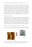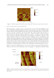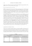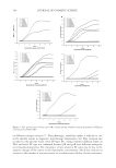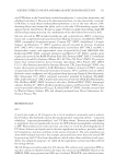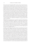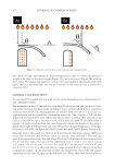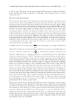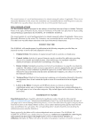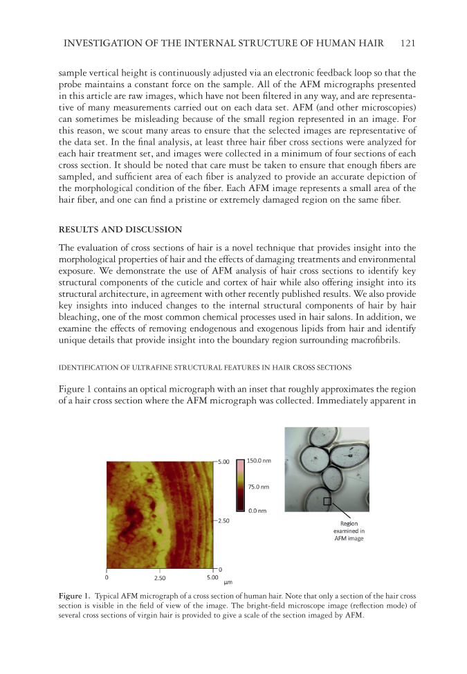
INVESTIGATION OF THE INTERNAL STRUCTURE OF HUMAN HAIR 121 sample vertical height is continuously adjusted via an electronic feedback loop so that the probe maintains a constant force on the sample. All of the AFM micrographs presented in this article are raw images, which have not been fi ltered in any way, and are representa- tive of many measurements carried out on each data set. AFM (and other microscopies) can sometimes be misleading because of the small region represented in an image. For this reason, we scout many areas to ensure that the selected images are representative of the data set. In the fi nal analysis, at least three hair fi ber cross sections were analyzed for each hair treatment set, and images were collected in a minimum of four sections of each cross section. It should be noted that care must be taken to ensure that enough fi bers are sampled, and suffi cient area of each fi ber is analyzed to provide an accurate depiction of the morphological condition of the fi ber. Each AFM image represents a small area of the hair fi ber, and one can fi nd a pristine or extremely damaged region on the same fi ber. RES ULTS AND DISCUSSION The evaluation of cross sections of hair is a novel tec hnique that provides insight into the morphological properties of hair and the effects of damaging treatments and environmental exposure. We demonstrate the use of AFM analysis of hair cross sections to identify key structural components of the cuticle and cortex of hair while also offering insight into its structural architecture, in agreement with other recently published results. We also provide key insights into induced changes to the internal structural components of hair by hair bleaching, one of the most common chemical processes used in hair salons. In addition, we examine the effects of removing endogenous and exogenous lipids from hair and identify unique details that provide insight into the boundary region surrounding macrofi brils. IDE NTIFICATION OF ULTRAFINE STRUCTURAL FEATURES IN HAIR CROSS SECTIONS Fig ure 1 contains an optical micrograph with an inset that roughly approximates the region of a hair cross section where the AFM micrograph was collected. Immediately apparent in Figu re 1. Typical AFM micrograph of a cross section of human hair. Note that only a section of the hair cross section is visible in the fi eld of view of the image. The bright-fi eld microscope image (refl ection mode) of several cross sections of virgin hair is provided to give a scale of the section imaged by AFM.
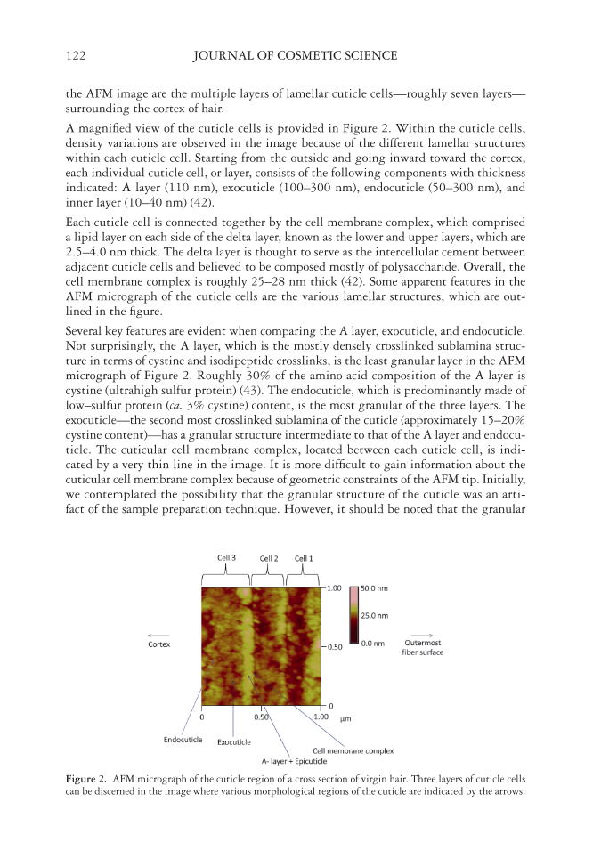
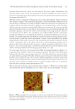
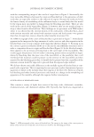
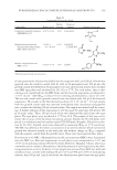
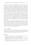
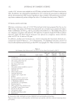
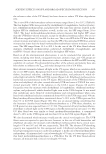
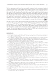
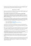
JOURNAL OF COSMETIC SCIENCE 122 the AFM image are the multiple layers of lamellar cuticle cells—roughly seven layers— surrounding the cortex of hair. A m agnifi ed view of the cuticle cells is provided in Figure 2. Within the cuticle cells, density variations are observed in the image because of the different lamellar structures within each cuticle cell. Starting from the outside and going inward toward the cortex, each individual cuticle cell, or layer, consists of the following components with thickness indicated: A layer (110 nm), exocuticle (100–300 nm), endocuticle (50–300 nm), and inner layer (10–40 nm) (42). Eac h cuticle cell is connected together by the cell membrane complex, which comprised a lipid layer on each side of the delta layer, known as the lower and upper layers, which are 2.5–4.0 nm thick. The delta layer is thought to serve as the intercellular cement between adjacent cuticle cells and believed to be composed mostly of polysaccharide. Overall, the cell membrane complex is roughly 25–28 nm thick (42). Some apparent features in the AFM micrograph of the cuticle cells are the various lamellar structures, which are out- lined in the fi gure. Sev eral key features are evident when comparing the A layer, exocuticle, and endocuticle. Not surprisingly, the A layer, which is the mostly densely crosslinked sublamina struc- ture in terms of cystine and isodipeptide crosslinks, is the least granular layer in the AFM micrograph of Figure 2. Roughly 30% of the amino acid composition of the A layer is cystine (ultrahigh sulfur protein) (43). The endocuticle, which is predominantly made of low–sulfur protein (ca. 3% cystine) content, is the most granular of the three layers. The exocuticle—the second most crosslinked sublamina of the cuticle (approximately 15–20% cystine content)—has a granular structure intermediate to that of the A layer and endocu- ticle. The cuticular cell membrane complex, located between each cuticle cell, is indi- cated by a very thin line in the image. It is more diffi cult to gain information about the cuticular cell membrane complex because of geometric constraints of the AFM tip. Initially, we contemplated the possibility that the granular structure of the cuticle was an arti- fact of the sample preparation technique. However, it should be noted that the granular Figu r e 2. AFM micrograph of the cuticle region of a cross section of virgin hair. Three layers of cuticle cells can be discerned in the image where various morphological regions of the cuticle are indicated by the arrows.
Purchased for the exclusive use of nofirst nolast (unknown)
From: SCC Media Library & Resource Center (library.scconline.org)