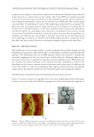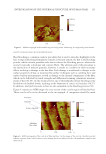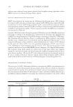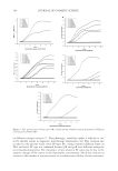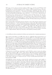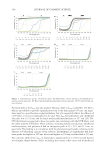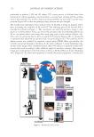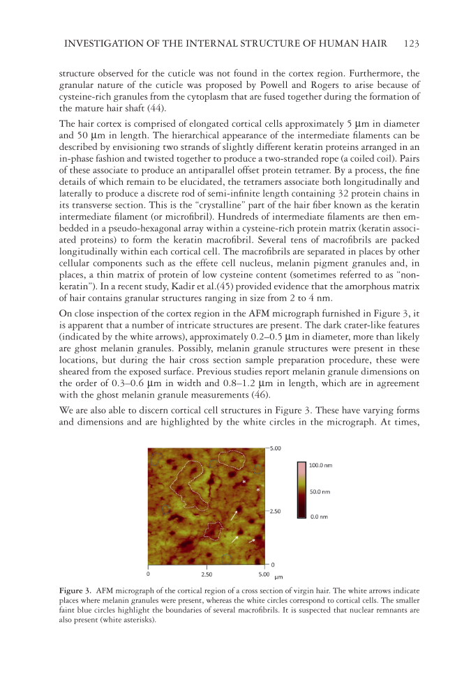
INVESTIGATION OF THE INTERNAL STRUCTURE OF HUMAN HAIR 123 structure observed for the cuticle was not found in the cortex region. Furthermore, the granular nature of the cuticle was proposed by Powell and Rogers to arise because of cysteine-rich granules from the cytoplasm that are fused together during the formation of the mature hair shaft (44). The hair cortex is comprised of elongated cortical cells approximately 5 μm in diameter and 50 μm in length. The hierarchical appearance of the intermediate fi laments can be described by envisioning two strands of slightly different keratin proteins arranged in an in-phase fashion and twisted together to produce a two-stranded rope (a coiled coil). Pairs of these associate to produce an antiparallel offset protein tetramer. By a process, the fi ne details of which remain to be elucidated, the tetramers associate both longitudinally and laterally to produce a discrete rod of semi-infi nite length containing 32 protein chains in its transverse section. This is the “crystalline” part of the hair fi ber known as the keratin intermediate fi lament (or microfi bril). Hundreds of intermediate fi laments are then em- bedded in a pseudo-hexagonal array within a cysteine-rich protein matrix (keratin associ- ated proteins) to form the keratin macrofi bril. Several tens of macrofi brils are packed longitudinally within each cortical cell. The macrofi brils are separated in places by other cellular components such as the effete cell nucleus, melanin pigment granules and, in places, a thin matrix of protein of low cysteine content (sometimes referred to as “non- keratin”). In a recent study, Kadir et al.(45) provided evidence that the amorphous matrix of hair contains granular structures ranging in size from 2 to 4 nm. On cl ose inspection of the cortex region in the AFM micrograph furnished in Figure 3, it is apparent that a number of intricate structures are present. The dark crater-like features (indicated by the white arrows), approximately 0.2–0.5 μm in diameter, more than likely are ghost melanin granules. Possibly, melanin granule structures were present in these locations, but during the hair cross section sample preparation procedure, these were sheared from the exposed surface. Previous studies report melanin granule dimensions on the order of 0.3–0.6 μm in width and 0.8–1.2 μm in length, which are in agreement with the ghost melanin granule measurements (46). We are also able to discern cortical cell structures in Figure 3. These have varying forms and dimensions and are highlighted by the white circles in the micrograph. At times, Figur e 3. AFM micrograph of the cortical region of a cross section of virgin hair. The white arrows indicate places where melanin granules were present, whereas the white circles correspond to cortical cells. The smaller faint blue circles highlight the boundaries of several macrofi brils. It is suspected that nuclear remnants are also present (white asterisks).

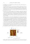
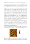
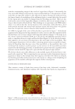
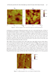
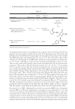
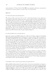
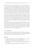
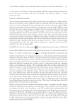
JOURNAL OF COSMETIC SCIENCE 124 cortical cells can have an irregular shape as illustrated in the fi gure. The small features in Figure 3, outlined in blue, correspond to macrofi brils. At a higher resolution, we can see the clear delineation between several macrofi brils (see Figure 4). Each macrofi bril, measured to be approximately 400 nm in diameter, is surrounded by a thin boundary. It has been suggested in the past that there is a demarcation between adjacent macrofi brils denoted as intermacrofi brillar material (47). Most reference texts report that the macrofi bril dimensions for human hair typically range from 40 to 500 nm in diameter (43,48,49). In wool, there are three types of cortical cells: paracortical, orthocortical, and mesocortical cells. In human hair, researchers have used terminology such as ortho-type and para-type cortical cells to denote the similarity of cell types that share some common features with those from wool. The size and struc- ture of macrofi brils in wool are fairly consistent for each cell type. However, the macrofi - brils in human hair were reported to vary considerably in size and shape (50). Therefore, it should come of no surprise that there is such high variability in the size of macrofi brils reported from various sources. Regardless, our measured values (approximately 400 nm) appear to be in line with those previously reported in the literature. Noteworthy, in a study carried out by Harland et al., (47) the thickness of the cross section was found to cause some variability in the measurement of macrofi brillar dimensions because of the tilt angle of intermediate fi laments. Another key feature of hair morphology is the medulla. It is not always present in hair fi bers and is most commonly found in hair of Asian descent. For example, in Caucasian hair, the medulla is not continuous along the length of the fi ber, so depending on the location from which the fi ber cross section is obtained, you may or may not observe the medulla structure. Regardless, Figure 5 contains an AFM image of the cross section of hair containing a medulla. In agreement with previous studies, the medulla appears as a porous and rough structure (51). Historically, not that much attention was given to understanding the structure and func- tion of the medulla. It was merely thought to be a vacuolated morphological component of the fi ber. Studies in recent years, however, have shown that it is rich in lipids and also contain fi brillar protein structures (35,51,52). Furthermore, studies have demonstrated that the medulla is a fairly complex structure and consists of a medullary tube surrounded by two types of medullary cells and vesicles with proteinaceous granules (53). Figure 4. AFM micrograph of a magnifi ed section of the region shown in Figure 3. The slightly elliptically shaped feature in the center of the image is a macrofi bril.
Purchased for the exclusive use of nofirst nolast (unknown)
From: SCC Media Library & Resource Center (library.scconline.org)