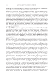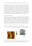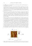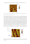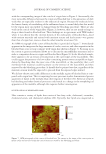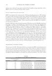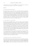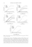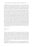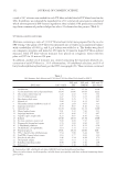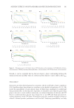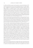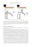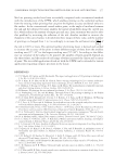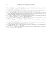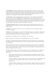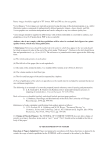
INVESTIGATION OF THE INTERNAL STRUCTURE OF HUMAN HAIR 127 endogenous or exogenous. Endogenous lipids carry out a structural function, serving as the building blocks of the cell membrane complex in hair. Compositionally and structur- ally, there are three different types of cell membrane complex in hair fi bers: (i) cuticle cell membrane complex situated above and below each cuticle cell, (ii) cortex cell membrane complex that surrounds each spindle-shaped cortical cell, and (iii) cuticle–cortex cell membrane complex that provides the boundary between the cuticle and cortex (61). Exogenous lipids arise from the sebaceous gland, which coats the exterior of the fi ber and also secretes lipids within the fi ber structure. The Soxhlet extraction method used in this study more than likely removes both endog- enous and exogenous lipids leaving only covalently bound 18-MEA, which is present in the outermost layer of each cuticle cell. Figure 9 contains an AFM micrograph of the cuticle of delipidated hair. It appears there is a greater degree of demarcation between cuticle cells than the virgin hair (see Figure 2). In addition, the cells adopt a swollen appearance—similar to that found in an earlier TEM study by Takizawa et al. (62) in permanent waved hair. The density of the various lamellar structures is less distinct in the delipidated hair than virgin and bleached hair. Overall, there appears to be a less granular Figure 8. A F M micrographs of the cortex of (A) virgin hair and (B) bleached hair. Figure 9. AF M micrograph of the cortical region of delipidated hair.
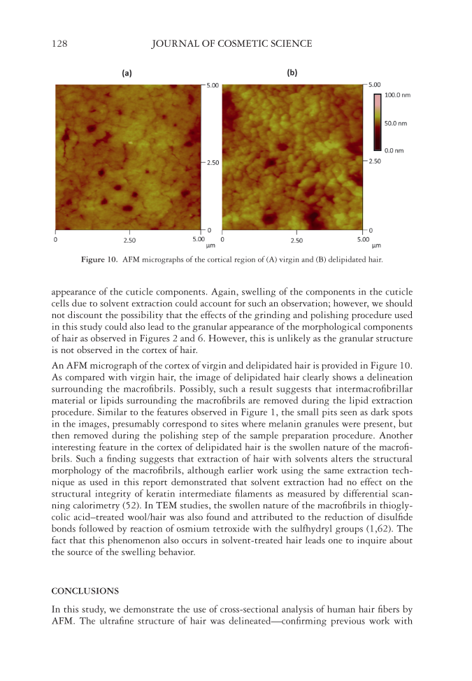
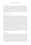
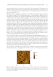
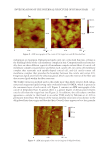
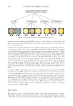
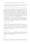
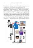
JOURNAL OF COSMETIC SCIENCE 128 appearance of the cuticle components. Again, swelling of the components in the cuticle cells due to solvent extraction could account for such an observation however, we should not discount the possibility that the effects of the grinding and polishing procedure used in this study could also lead to the granular appearance of the morphological components of hair as observed in Figures 2 and 6. However, this is unlikely as the granular structure is not observed in the cortex of hair. An AFM micrograph of the cortex of virgin and delipidated hair is provided in Figure 10. As compared with virgin hair, the image of delipidated hair clearly shows a delineation surrounding the macrofi brils. Possibly, such a result suggests that intermacrofi brillar material or lipids surrounding the macrofi brils are removed during the lipid extraction procedure. Similar to the features observed in Figure 1, the small pits seen as dark spots in the images, presumably correspond to sites where melanin granules were present, but then removed during the polishing step of the sample preparation procedure. Another interesting feature in the cortex of delipidated hair is the swollen nature of the macrofi - brils. Such a fi nding suggests that extraction of hair with solvents alters the structural morphology of the macrofi brils, although earlier work using the same extraction tech- nique as used in this report demonstrated that solvent extraction had no effect on the structural integrity of keratin intermediate fi laments as measured by differential scan- ning calorimetry (52). In TEM studies, the swollen nature of the macrofi brils in thiogly- colic acid–treated wool/hair was also found and attributed to the reduction of disulfi de bonds followed by reaction of osmium tetroxide with the sulfhydryl groups (1,62). The fact that this phenomenon also occurs in solvent-treated hair leads one to inquire about the source of the swelling behavior. CONCLUSIONS In this study, we demonstrate the use of cross-sectional analysis of human hair fi bers by AFM. The ultrafi ne structure of hair was delineated—confi rming previous work with Figure 10. AF M micrographs of the cortical region of (A) virgin and (B) delipidated hair.
Purchased for the exclusive use of nofirst nolast (unknown)
From: SCC Media Library & Resource Center (library.scconline.org)