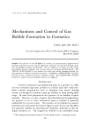
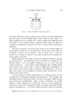
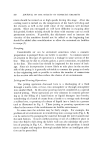
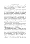
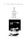
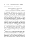
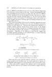
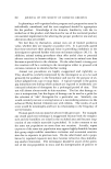
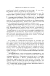
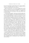
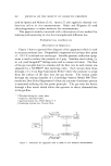
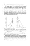
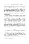
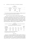
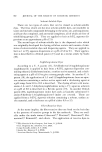
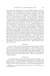
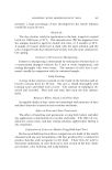
362 JOURNAL OF THE SOCIETY OF COSMETIC CHEMISTS used by Spruit and Malten (5, 6). Spruit (7) also applied a thermal con- ductivity cell to in vivo measurements. Baker and Kligman (8) used electrohygrometry to make moisture loss measurements. This paper is mainly concerned with a description of our method for isolating and measuring in vivo the transepidermal diffusion loss. EXPERIMENTAL AND R•ESULTS Description of Apparatus Figure 1 shows a general flow diagram of the apparattis which is used to measure moisture loss. Prepurified compressed air having a dew point of --59.5øC is utilized as a carrier gas. Suitable pressure reduction equip- ment is used to reduce the pressure to 5 psig. Stainless steel tubing (•/• in. o.d.) and Swagelok '©• fittings were used to connect all units. The flow of the gas was split into two streams and the flow rate in each stream was adjusted by a NUPRO ©* fine metering valve. Each stream then flows through a 4 X 4 cm Sage Instrument Probe* where the moisture is swept from the surface of the skin into the gas stream. The stream passes through the sensing chamber of a Cambridge Systems Model 990 Ther- moelectric Dew Point HygTometerõ where the amount of moisture present is measured utilizing the dew point principle. The gas exits the device through a flow meter which allows the operator to detect abnormalities in the gas flow. Crawford Fitting Co., Solon, Ohio. Nupro Co., Cleveland, Ohio. Sage Instruments, Inc., White Plains, N.Y. Cambridge Systems, Inc., Newton, Mass. INSTRUMENT , Figure 1. Schematic for apparatus used to make in vivo moisture measurements

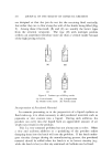
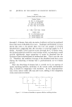
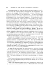
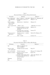
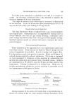
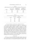
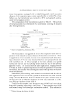
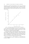
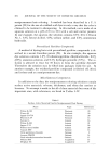
TRANSEPIDERMAL MOISTURE LOSS 363 Each dew point instrument is attached to one side of a two-pen re- corder. An electronic calibration unit is also attached to equalize the electrical response of the two instruments. The range of moisture levels which may be measured is determined by the flow rate. A rate of 50 ml/min allows measurements of 0 to 10 mg/cm2hr with a 50% scale reading of 0.3 mg/cm2hr. Standardization of Apparatus The Sage Instrument Probe is replaced with a gas chromatographic type injector port. Standardization is accomplished by leaking water at several different rates into the gas stream utilizing a radiometer auto- matic syringe microburet fitted with a 2-v1 gas chromatog•:aphic syringe. Standard response graphs are plotted and used to interpret the instru- mental response. Environmental Precautions Water measured by the apparatus in in vivo experiments could come from three sources. These sources are listed in Table I along with the orders of magnitude of each effect. Eccrine sweating is the largest effect. It is discontinuous and varies greatly in amount. Baker and Kligman (8) utilized pharmacologically induced anhydrosis. Not being so equipped we have attempted to eliminate eccrine sweating by maintaining the thermal and emotional environment below threshold values. Subjects are conditioned to a 19.4-20.0øC room for 20 min and then tested at this temperature. Testing is conducted in an isolated area. While not ab- solutely guaranteeing anhydrosis, eccrine sweating is controlled out of our measurement within practical limits. Table I Basic Mechanisms by which Water Is Lost through Skin Rate Mechanism (mg/cm2/hr) Duration Eccrine sweating Transepidermal diffusion Stratum corneum desorption of normal hydrated skin by a stream of dry gas 32-48 Discontinuous 0.2-0.3 Continuous 0.6-0.7 10-15 rain Stratum Comeurn Desorption During exposure of the surface of normal skin to the dehydrating in- fluenee of a stream of dry air, some stratum comeurn desorption of the
Purchased for the exclusive use of nofirst nolast (unknown)
From: SCC Media Library & Resource Center (library.scconline.org)