j. Soc. Cosmet. Chem., 43, 215-217 (July/August 1992) Iron content of human epidermis from sun-exposed and non-exposed body sites DONALD L. BISSETT and JAMES F. McBRIDE, The Procter & Gamble Company, Miami Valley Laboratories, Cindnnati, OH 45239-8707. Received March 3, 1992. Synopsis In previous work we observed that the iron content of human dermis is greater in sun-exposed than in non-exposed body sites. The present study indicates a similar difference in iron content for epidermis from these body sites. We speculate on the possible role of the iron in epidermal photodamage. INTRODUCTION Chronic exposure of hairless mice to UVB (1) or UVC (2) has been shown to result in an increase in non-heme iron content of the exposed skin. Analysis of human skin from sun-exposed vs non-exposed body sites also revealed a greater content of iron in the former (1). Based on histological and immunohistochemical analyses (1), the iron in UV-exposed mouse and human skins appeared to be present primarily as large dermal deposits. Iron can participate in generation of oxygen radicals (3-6), which are dam- aging to biological targets such as protein, lipid, and nucleic acid (6-11). The increased iron in UV-exposed skin would increase the likelihood of damage via this mechanism. The observation that topical iron chelators are photoprotective in mice (1) suggests that this mechanism is a significant factor in photodamage. Since topical chelators were protective against damage to both dermal and epidermal components (1), it is likely that substantial levels of iron are present in the epidermis, as they are in the dermis. In the present study, iron analysis of human epidermis was done to measure the basal level of this element in skin from non-exposed sites and, as was observed previously for the dermis, to determine if the content is greater in sun- exposed sites. MATERIALS AND METHODS HUMAN EPIDERMIS Human skin was obtained from remaining frozen samples of the same donor skins described previously (1). Donors were two males and six females, ages 53 to 80 years. 215
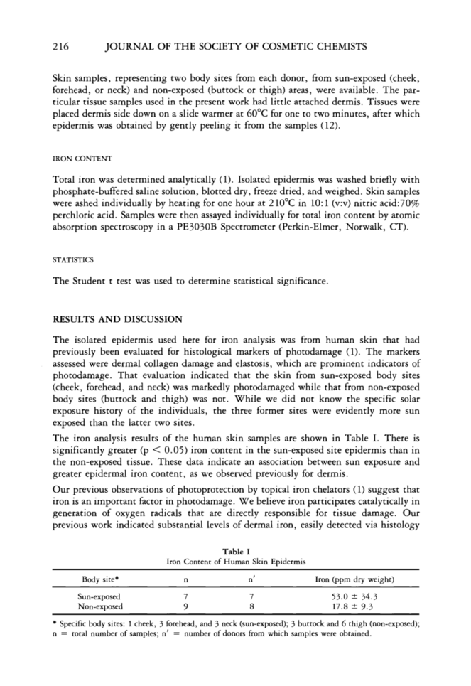
216 JOURNAL OF THE SOCIETY OF COSMETIC CHEMISTS Skin samples, representing two body sites from each donor, from sun-exposed (cheek, forehead, or neck) and non-exposed (buttock or thigh) areas, were available. The par- ticular tissue samples used in the present work had little attached dermis. Tissues were placed dermis side down on a slide warmer at 60øC for one to two minutes, after which epidermis was obtained by gently peeling it from the samples (12). IRON CONTENT Total iron was determined analytically (1). Isolated epidermis was washed briefly with phosphate-buffered saline solution, blotted dry, freeze dried, and weighed. Skin samples were ashed individually by heating for one hour at 210øC in 10:1 (v:v) nitric acid:70% perchloric acid. Samples were then assayed individually for total iron content by atomic absorption spectroscopy in a PE3030B Spectrometer (Perkin-Elmer, Norwalk, CT). STATISTICS The Student t test was used to determine statistical significance. RESULTS AND DISCUSSION The isolated epidermis used here for iron analysis was from human skin that had previously been evaluated for histological markers of photodamage (1). The markers assessed were dermal collagen damage and elastosis, which are prominent indicators of photodamage. That evaluation indicated that the skin from sun-exposed body sites (cheek, forehead, and neck) was markedly photodamaged while that from non-exposed body sites (buttock and thigh) was not. While we did not know the specific solar exposure history of the individuals, the three former sites were evidently more sun exposed than the latter two sites. The iron analysis results of the human skin samples are shown in Table I. There is significantly greater (p 0.05) iron content in the sun-exposed site epidermis than in the non-exposed tissue. These data indicate an association between sun exposure and greater epidermal iron content, as we observed previously for dermis. Our previous observations of photoprotection by topical iron chelators (1) suggest that iron is an important factor in photodamage. We believe iron participates catalytically in generation of oxygen radicals that are directly responsible for tissue damage. Our previous work indicated substantial levels of dermal iron, easily detected via histology Table I Iron Content of Human Skin Epidermis Body site* n n' Iron (ppm dry weight) Sun-exposed 7 7 53.0 -+ 34.3 Non-exposed 9 8 17.8 + 9.3 * Specific body sites: 1 cheek, 3 forehead, and 3 neck (sun-exposed) 3 buttock and 6 thigh (non-exposed) n = total number of samples n' = number of donors from which samples were obtained.
Purchased for the exclusive use of nofirst nolast (unknown)
From: SCC Media Library & Resource Center (library.scconline.org)


