192 .JOURNAL OF THE SOCIETY OF COSMETIC CHEMISTS Cocci were classified in accordance with the Baird-Parker (11) system of classification. The organisms conformed to the characteristics of Staphylococcus subgroup II. Growth was in Brain Heart Infusion (Difco) medium at g7øC with the cells harvested by centrifugation after 48 hours of incubation. l Inless otherwise specified, organisms were added to artificial sebum at a concentration of 100 mg/g. The microbial concentrate was ob- tained by centrifuging growth medium at 10,000 rpm in a refrigerated centrifuge for 20 min. The spent medium was decanted and the cen- trifuged cells were then weighed and added directly and with vigorous stirring to the artificial sebum. This mixture was then applied to the delineated areas on the dorsal surface of the guinea pig. Fatty A cid Analyses Extractions of guinea pig skin were carried out by placing a glass collar 25 mm in diameter firmly on the test site. A 5-ml volume of 4:1 diethyl ether:methanol was placed within the collar and the area was scrubbed for 30 sec with a glass rod flattened at the tip. The extract was then withdrawn and filtered and the constitttent free fatty acids were converted to the corresponding methyl esters by the methods of Metcalf et al. (12) Gas chromatography was carried out with an FgcM model 720 dual column gas chromatograph* utilizing a thermal conductivity de- tector. A 10-foot stainless steel column of l/4-in. diameter, packed with a 1:9 mixture of diethylene glycol succinate* and 60/80 mesh Gas Chrom P,* and operating at a temperature of 185 ø or 190øC, was used in these studies. RESULTS Early experiments had shown that the addition of buffered suspen- sions of scalp microorganisms directly to guinea pig skin produced no visible irritation or other reaction. In view of the high concentrations of fatty materials on the human scalp and the relatively low concentra- tions of lipid on guinea pig skin, an attempt was made to approximate more closely the conditions existing on the httman scalp by supplement- ing the guinea pig skin lipids with the artificial sebum for•nulation. The various strains of scalp organisms were then admixed with the at- * F & M Scientific Corp., Avondale, Pa. ? Analytical Engineering Laboratories, Hamden, Co•m. • Applied Sciences I•aboratories, State College, Pa.

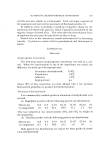
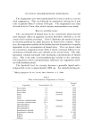
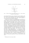
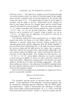
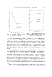
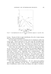
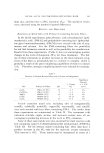
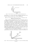
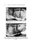
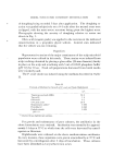
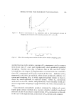
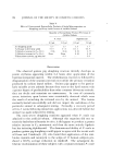
MOI)EL SYSTEM FOR DANDRUFF INVESTIGATION Table III Effcct of Cell Suspensions on the Initiation of Guinea Pig Sloughing in the Presence and Absence of Artificial Sebum 193 Sloughing Reaction Antibiotic s Treated No Antibiotic P. ovale q- saline P. ovale q- artificial sebum Staphylococcus JTM q- saline Staphylococcus JTM q- artificial sebum Diphtheroid q- saline Diphtheroid q- artificial sebum Control, artificial sebum 0 0 3 3 0 0 1 0 0 0 2 2 0 0 • O. 05% solution of tetracycline. tificial sebum before application to the guinea pig. The results (Table IIl) show that suppression of the natural flora of the guinea pig skin does not appear necessary for a positive test reaction and that concen- trated P. ovale cells produced the strongest reaction of the three groups of microorganisms tested. The most important conclusion from these data is that the artificial sebum must be present before the microorganisms tested can induce sloughing. The sloughing reaction appeared within 4 to 5 days and reached maximal severity within 6 to 7 days. Under normal circum- stances the reaction began to subside within an additional 10 days, with total clearing normally occurring within 2 to 3 weeks from the time of initial application. The scales observed were large, waxy segments of stratum corneum resembling human dandruff to the unaided eye. Microscopically, the guinea pig skin scales bore a remarkable resemblance to human dandruff scales. Thickened, horny, keratin layers were ob- served in both human dandruff and the sloughed guinea pig skin flakes. Attempts to accelerate and/or accentuate the sloughing reaction by preabrading the skin or applying the irritant mixture to other areas of the guinea pig were unsuccessful. Koch's postulates have been fulfilled repeatedly using either the P. ovale or diphtheroid cell suspensions in artificial sebum. There were no obvious differences in micro- or macro- scopic appearance traceable to the type of inoculum used. The relative ability of representative scalp microorganisms to produce sloughing is shown in Fig. 2. It is apparent that the staphylococcus was unable to produce appreciable sloughing with even very high cell con- centrations. The suspension of P. ovaIe was the most effective in this
Purchased for the exclusive use of nofirst nolast (unknown)
From: SCC Media Library & Resource Center (library.scconline.org)