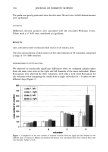
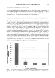
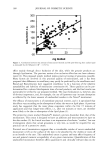
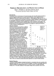
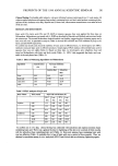
UREA ANALYSIS OF STRATUM CORNEUM 163 5% Urea increases significantly the urea content of stratum corneum compared to an untreated area and a treatment with Laceran © Spezial Creme, respectively. The increased urea amount remains at a high level for at least 24 hours after final application. This lasting effect is a possible factor in the increased hydration of skin surface following treatment with the urea-containing product compared with the non-urea-containing control product. We would propose that the reduced amount of urea in skin extracts of skin areas treated with Laceran © Spezial Creme is caused by reduced extractability of urea as a consequence of a visible residual lipid layer even after 24 hours following final application. This, however, is not found for the Laceran © Spezial Creme 5 % Urea, which differs in basis formulation from the Laceran © Spezial Creme. Clearly, urea treatment of skin causes an increase of skin hydration as measured by corneometry. This enhancement results in an improved clinical appearance, i.e., smooth- ness, scaliness, or erythema, of patients with dry or xerotic skin (5, 6, 16, 17). Our results support the concept of treatment of skin diseases, e.g., atopic dermatitis, or conditions of clinical dry skin with urea-containing products to compensate for the reduced amount of urea in stratum corneum (1). REFERENCES (1) W. Raab, Harnstoff in der Dermatologie, TIV Dermatol,, 23, 257-269 (1993). (2) W. Wohlrab, Zur Verwendung von Harnstoff in der Dermatologie, Dr. Derm., 36, 528-537 (1988). (3) H.W. Spier and C. Carrie, Hornschichtphysiologie als gewerbedermatologische Grundlagenfbrsch- ung, BerujS-Dermatosen, 15, 121-146 (1967). (4) G. Swanbeck and G. Rajka, Antipruritic effect of urea solutions, Acta Derm. Venereol. (Stockholm), 50, 225-227 (1970). (5) J. Serup, A double-blind comparison of two creams containing urea as the active ingredient, Acta Derm. Venereol. (Stockholm), Suppl. 177, 34-38 (1992). (6) J. Serup, A three-hour test for rapid comparison of effects of moisturizers and active constituents (urea), Acta Derm. Venereol. (Stockholm), Suppl, 177, 29-33 (1992). (7) H. W. Spier and G. Pascher, Die wasserli3slichen Bestandteile der peripheren Hornschicht (Hauto- berfiiiche), Arch. Dermatol. Syph., 199, 411-427 (1955). (8) O. Jacobi, Die Inhaltsstoffe des normalen Stratum corneum und Callus menschlicher Haut, Arch. Derm. Fo,xch., 240, 107-118 (1971). (9) L. Kerscher and J. Ziegenhorn, "Urea" in MethodJ of Enzymatic Analysis, 3rd ed., J. Bergmeyer and M. GraB1, Eds. (VCH-Verlagsgesellschaft mbH, Weinheim, 1985), Vol. 8, pp. 444-453. (10) E. Schwarz, Freie Aminosiiuren und verwandte Verbindungen in der abschabbaren Hornschicht un- befallener Haut von mikrobiellen Ekzematikern und Hautgesunden, Arch. Derre. Forsch., 242, 87-96 (1971). (11) E. Schwarz, Biochemische Stigmata menschlicher Hautoberfi•/che im Alter, Z. Klin. Chem. Klin. Blochim., 12, 93-97 (1974). (12) E. Schwarz, Neurodermitis und Hornschicht, Z. Hautk., 52 (Suppl. 2), 59-64 (1977). (13) G. Kloss and E. Schwarz, Freie Aminos•/uren und andere Ninhydrin-positive Substanzen in normalet Hornschicht und in Psoriasis-Schuppen, Arch. Klin. Exp. Dermatol., 228, 188-198 (1967). (14) J. Koyama, I. Horii, K. Kawasaki, Y. Nakayama, Y. Morikawa, and T. Mitsui, Free amino acids of stratum corneum as a biochemical marker to evaluate dry skin, J. Soc. Cosmet. Chem., 35, 183-195 (1984). (15) J. Ennen and D. Hiintschel, unpublished data (1997). (16) G. Swanbeck, Urea in the treatment of dry skin, Acta Derm. Venereol. (Stockholm), Suppl. 177, 7-8 (1992). (17) P.D. Pigatto, A. S. Bigardi, C. Cannistraci, and M. Picatrio, 10% urea cream (Laceran) for atopic dermatitis: A clinical and laboratory evaluation, J. Dermatol. Treat., 7, 171-145 (1996).

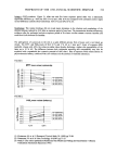
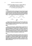
j. Cosmet. Sci., 49, 165-174 (May/June 1998) Ouantification of depletion in solution-type topical preparations in vivo CLAUDIA S. LEOPOLD, Department of Pharmacez/tical Strafie Technology, Heinrich Heine University, Universiti/tsstrasse 1, 40225 Di/sseldorf Germany. Accepted for publication May 15, 1998. Part of this paper was presented as a poster at the AAPS Annual Meeting and Exposition, Miami Beach, FL, November 5-9, 1995. Synopsis To distinguish between thermodynamic, penetration-enhancing, and permeant depletion effects of solution- type transdermal preparations, two sets of human in vivo permeant penetration data obtained under finite- dose and infinite-dose conditions, respectively, were compared. The measurement of the pharmacodynamic response of a permeant or the determination of permeant penetration rates under finite-dose conditions includes all three types of vehicle effects, whereas under infinite-dose conditions permeant depletion does not play a significant role. Four lipophilic liquids (caprylic/capric triglyceride, isopropyl myristate, light mineral oil, and dimethicone 100) were used as vehicles. The reciprocal of the time of onset of an erythema (1/LT) and the duration of the erythema D induced by the model compound methyl nicotinate served as response parameters. Steady-state penetration rate measurements were made with a recently developed glass chamber system. Bioavailability factors f and enhancement factors EF were determined from both methyl nicotinate penetration rates (infinite-dose conditions) and the horizontal distances between concentration- response curves (finite-dose conditions), with caprylic/capric triglyceride as the standard vehicle. Permeant depletion was quantified by dividing the enhancement factors determined under infinite-dose conditions by those under finite-dose conditions. These ratios were called depletion factors DF. Significant methyl nico- tinate depletion was observed with both response parameters. However, the most accurate bioavailability factors may be obtained with the response parameter 1/LT, especially if they are determined from the horizontal distances between the curves in the high-response region where parallelism is given. INTRODUCTION With solution-type permeant preparations applied to the skin, three types of penetra- tion-influencing effects may be observed: (a) thermodynamic effects resulting from different permeant solubilities in the vehicles, (b) penetration-enhancing effects caused by an interaction of the vehicle with the barrier stratum corneum, and (c) permeant depletion in the vehicle in the case of finite-dose conditions. Permeant depletion may result from a high thermodynamic activity of the permeant in the vehicle and/or from pronounced penetration enhancement caused by vehicle com- ponents or by the vehicle itself. The extent of this effect depends on the thickness of the
Purchased for the exclusive use of nofirst nolast (unknown)
From: SCC Media Library & Resource Center (library.scconline.org)