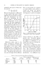
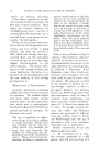
36 JOURNAL OF THE SOCIETY OF COSMETIC CHEMISTS alanine and not on tyrosine (Bloch and Schaaf (7) and our own experi- .ments). The polyphenol oxidase of the myelogenic cells acts on many poly- phenol derivatives and easily oxi- dizable substances such as epi- nephrine, pyrogallol and phenylene diamine (Bloch and Peck (1{5)) as well as on dioxyphenylalanine. In view of the higher specificity of the dioxyphenylalanine oxidase of the melanoblasts, any theory attempt- ing to identify dioxyphenylalanine oxidase with polyphenolase (Oppen- heimer (17)) is just as untenable as that of the identity of dioxyphenyl- alanine oxidase with tyrosinase. There are a number of proofs for the existence of an individual di- oxyphenylalanine enzyme: (1) The dioxyphenylalanine oxidase is ex- tremely labile against the destruc- tive influences of heat and especially of certain poisons as compared to the less specific, more rugged poly- phenolase and the nonenzymatic oxidizing catalysts of the cell. (2) Its action is confined to a much narrower hydrogen ion concentra- tion than that of either polypheno- lase or tyrosinase. (3) As demon- strated by Bloch and Schaaf (7), the dioxyphenylalanine oxidase is so specific as to be inac'tive against a substrate showing the slightest change in the chemical constitution. Recently Mulzer and Schmalfuss (18) cast doubt on the specificity of the reaction to dioxyphenylalanine because in their hands 3-4 dioxy- phenylethylamine (oxytramine) also gave a positive product of dioxy- phenylalanine, it seems that this finding rather corroborates Bloch's theory of the formation of pigment. In the following series of experi- ments new evidence is presented for the specificity of the reaction to dioxyphenylalanine. The optical specificity of the action of enzymes has been studied thoroughly in several groups of hydrolyzingenzymes. The problem of optical specificity in oxidizing enzymes has been investigated with tyrosinase. According to Abderhalden and Guggenheim (19), this enzyme is optically selective to a limited degree only, showing some preference for the leveroratory natu- ral substrate. We (Peck, Sobotka and Kahn (20)) investigated the optical speci- ficity of the dioxyphenylalanine oxidase, and we found that it was strictly specific for the levorotatory natural form of dioxyphenylalanine (21). The dextrorotatory antipode was left untouched by the enzyme (22). Bloch and Schaaf (7) have independently made the same ob- servation. The formation of melanin from dioxyphenylalanine by crude potato tyrosinase (prepared according to Lichtman and Sobotka (23)) is rapid. It takes place to a con- siderable degree even when the true tyrosinase is destroyed by heat. The potato preparation acts equally on dextrorotatory and on levo- rotatory dioxyphenylalanine, as is to be expected in a material rich in polyphenol-oxidizi.ne catalysts. The enzymes responsible for the

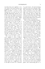
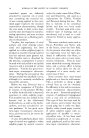
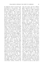
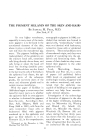
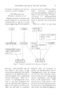
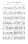
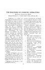
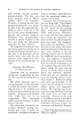
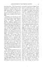
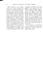
THE PIGMENT MELANIN OF THE SKIN AND HAIR 37 formation of melanin may be sum- marized as shown in Figure I. T•iE MEL^NOBL^STS (])IGMENT-FORMING CELLS) Pigment (melanin') is found in the human epidermis in the basal and overlying cells as well as in a more bizarre appearing cell with dendritic processes such as condylomas, lichen ruber, moll uscum contagiosun psoriasis and penphigus vegetans, as well as in the early stages of melano- carcinomas. In hyperpigmented and acanthotic process they may be found in the layers above the basal cells. While as a rule it is true that TYRC•IN[.] I DIOXYPHENYLALANINE 10XYTY DIOXYPHENYLALANINE OXIDASE HICHLY OPTICALLY•SPECIFIC • pH 7.3-7.5 • . .• J MELANIN TYRAMINE MONOPHENOLS'] e.g.,/:-CRESOL TYROSINASE - i,•ON 0 ?H ENOLASE OPTICALLY SPECIFIC: pH 6-8 RAMINE__ j DIPliENOLS '- I EPINEPHRINJ •"x-- e.g., CATECHOL POLYPHENOL/ • NONSPECIFIC - I) [1__•• Figure 1.---Enzymes responsible for the formation of melanin processes. Such dendritic cells are aisc) found in the matrix of pig- mented hairs, epidermal hair sheets, sometimes in the epidermal parts of sebaceous glands and in pigmented mucous membranes. They are in- creased in hyperpigmented proc- esses at the time of pigment for- mation, especially after exposure to light, x-rays, radium and thorium X. They are well seen in acanthotic dendritic cells are' increased at number and therefore better seen in hyperpigmented skins, for reasons not yet clear they are better visual- ized in acanthoses and in pigmented mucous membranes, even though there is little pigmentation. This may be due to some extent to the fact that the dendritic processes may be obscured by adjoining pigmented cells, and in the relatively pigment-
Purchased for the exclusive use of nofirst nolast (unknown)
From: SCC Media Library & Resource Center (library.scconline.org)