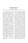
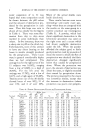
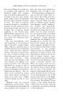
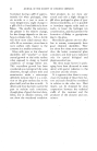
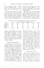
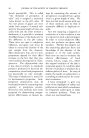
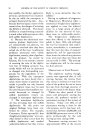
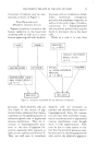
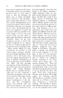
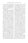
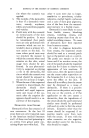
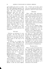
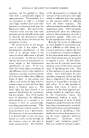
THE PIGMENT MELANIN OF THE SKIN AND HAIR 37 formation of melanin may be sum- marized as shown in Figure I. T•iE MEL^NOBL^STS (])IGMENT-FORMING CELLS) Pigment (melanin') is found in the human epidermis in the basal and overlying cells as well as in a more bizarre appearing cell with dendritic processes such as condylomas, lichen ruber, moll uscum contagiosun psoriasis and penphigus vegetans, as well as in the early stages of melano- carcinomas. In hyperpigmented and acanthotic process they may be found in the layers above the basal cells. While as a rule it is true that TYRC•IN[.] I DIOXYPHENYLALANINE 10XYTY DIOXYPHENYLALANINE OXIDASE HICHLY OPTICALLY•SPECIFIC • pH 7.3-7.5 • . .• J MELANIN TYRAMINE MONOPHENOLS'] e.g.,/:-CRESOL TYROSINASE - i,•ON 0 ?H ENOLASE OPTICALLY SPECIFIC: pH 6-8 RAMINE__ j DIPliENOLS '- I EPINEPHRINJ •"x-- e.g., CATECHOL POLYPHENOL/ • NONSPECIFIC - I) [1__•• Figure 1.---Enzymes responsible for the formation of melanin processes. Such dendritic cells are aisc) found in the matrix of pig- mented hairs, epidermal hair sheets, sometimes in the epidermal parts of sebaceous glands and in pigmented mucous membranes. They are in- creased in hyperpigmented proc- esses at the time of pigment for- mation, especially after exposure to light, x-rays, radium and thorium X. They are well seen in acanthotic dendritic cells are' increased at number and therefore better seen in hyperpigmented skins, for reasons not yet clear they are better visual- ized in acanthoses and in pigmented mucous membranes, even though there is little pigmentation. This may be due to some extent to the fact that the dendritic processes may be obscured by adjoining pigmented cells, and in the relatively pigment-

38 JOURNAL OF THE SOCIETY poor mucous membrane this .factor is obviated or that, as in acanthotic processes, there is a looser type of tissue so that the processes are better seen or better developed. If one examines a skin showing a fairly strong dopa reaction or one that is fairly well pigmented, one is struck by the clarity and the number of dendritic cells, seen in arete peg cut on a bias. It is possible that in many cases dendrites are given off in a plane which runs parallel to the body surface, so that in the usual section which cuts this plane at right angles the dendrites are poorly visualized. Above all, the state of pigment activity as well as the method employed for its demon- stration are of prime importance in the demonstration of dendritic cells. Many workers (Bloch [7]) have investigated the • problem of the nature, origin, anatomy, distribu- tion and biologic significance of the dendritic cell in the human skin, since the original demonstration of them in syphilitic leukoderma and in the normal hair matrix by Riehl (24) (1884). The first thorough investigation of their distribution in the normal white skin was made by Adachi (2'5) (1903). Recently Becker (26), in a detailed and thor- ough piece of work, undertook a systematic investigation of the pig- ment (melanin) of the upper mucous membranes and the skin, with special consideration of the den- dritic cells. His investigations showed that dendritic cells were much more frequent in the normal skin and mucous membranes than OF COSMETIC CHEMISTS has been supposed that they were found in all regions examined-- nipple, abdomen, axilla, toe, pre- puce, mouth, pharynx and esopha- gous--and that they varied in size, shape, nature of branching and numerical relation to nondendritic cells in different regions of the skin and mucous membranes and even in the same location in different per- sons. The dendritic cell in its appearance and variable form may appear very different from the ordi- nary basal cell. However, all tran- sitions between the most bizarre shapes with long, gnarled branches to basal cells with short stubby proc- esses can be seen (transition forms). The cell body is tisually larger than that of a basal cell and may be globular, irregularly oval, club- shaped or polyhedral. The proto- ß plasm is spongy, lacks fibrillation and contains pigment in its cell body as well as in the processes. The processes may be fine and whip- like, or thicker and irregularly formed, extending in the inter- cellular .spaces ahnost up to the stratum corneum (27). They may run along just below the basal cells, extending under three or four cells, and often curving upward at the end to go up between the basal cells again. From such a long process branches may be given off at right angles, also extending upward. The branches of a dendritic process often end in a Y-form or show at their terminations a button-like swelling. This knoblike or button- like swelling at the end of a process can be seen especially well in dopa
Purchased for the exclusive use of nofirst nolast (unknown)
From: SCC Media Library & Resource Center (library.scconline.org)