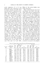
THE PIGMENT MELANIN OF THE SKIN AND HAIR 41 cutis waste elements or formed prod- ucts. This function, however, has not been proved. In the more recent papers of Masson (37), the dendritic cells have been classified as nerve cells and have been made identical with the Langerhans cells. A great deal of confusion has arisen because of the nomenclature used by various authors in writing about dendridc cells. In compari- son with the pigment-forming cells of the lower animals they have been called chromatophores or melano- phores. Still greater misunder- standing has been caused by their being called Langerhans cells (Masson (28 and 37), Pautrier, I,evy and Diss (35), Caudiere (36)), which are epidermal nerve elements and are not melanoblasts. As was shown by Bloch (7), quite apart from the question whether the den- dritic cells are closely allied or have their origin from nervous elements, they cannot be identified with the I•angerhans cells. This point will be taken up later. The terms in the following studies are those used by the Bloch school i.e., melanoblasts, pigment-building cells of the epi- dermis of dendritic or nondendritic forin and chromatophores, cells found in the cutis which phago- cytose pigment and are not pigment builders. The last-n, amed cells never show a positive dopa reaction, in contradistinction to the melano- blasts of mesodermal origin, which may also be found in the cutis in such conditions as blue nevus and mongolian spots. [)EI'IGMENrFATION BY •PECIFIC AN•I IOXIDANTS It was observed by Oettel (3S) in 1936 that the peroral actnfinistration of hydroquinone to black-haired cats turned the hair gray. Discon- tinuation of the drug resulted in repigmentation of the hair. In an investigation of occupational leuko- derma by Oliver, Schwartz and Warren (39 and 40), Schwartz found that the monobenzyl ether of hydroquinone, contained as an anti- oxidant in the rubber gloves of the workers, was responsible for the depigmentation. Their experi- ments as well as our own demon- strated that the alepigmentation was due to the action of.monobenzyl hydroquinone on the system "dopa"-oxidase dihydroxyphenyl- alanine ("dopa") (4l). Extensive experiments to study the mechanism of this depigmen- tation were carried out by Peck and Sobotka. (41). In a number of human subjects, both white and colored, the monobenzyl compound was applied in the form of a 50 per cent ointmentor as a 50 per cent ethereal suspension. In a few instances the concentrated powder was used. Leukoderma was pro- duced after incubation periods w•ry- ing from weeks. to months. Histologic examination of the areas of alepigmentation revealed a negative "dopa" reaction and an almost complete disappearance of the melanin. The microscopic pic- ture could not be differentiated from a vitiligo in many instances since the depigmentation was seen to

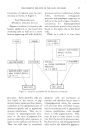
•2 JOURNAL OF THE SOCIETY OF COSMETIC CHEMISTS occur without preceding inflamma- tory reaction. A number of guinea pigs were fed approximately 12 grams of mono- benzyl ether of hydroquinone over a period of 5 months. This was well tolerated but no pigmentary changes were observed. Local application over a period of months, just as in the human subjects, caused de- pigmentation. The disappearance of the pigment was accompanied by a negative "dopa" reaction in the affected areas. This observation only applied to the previously pig- mented epidermis whereas the hair bulbs were not affected. This was probably due to a lack ofp4netration of the monobenzyl compound to the melanoblasts in the hair matrix. CAN PIGMENT ENZYME ACTIVITY BE STIMULATED IN UNPIGMENTED SKIN .• (42) The results of our experiments (42) have confirmed the observa- tions of Saxton, S•hmekebier and Kelley (43), as well as the earlier work of Loeb (44), Carnot and Deftandre (45) that the transplanta- tion of a black graft into a white skin resulted in the "extension" of the pigment into the surrounding un- pigmented host area while a white graft transplanted in a black skin ß was "invaded" by pigment from the ß host area. It was perfectly obvious from our histologic studies that the pigment formation in the white host area as well as in the non- pigmented grafts in no way differed from the ordinary mechanism of pig- ment formation. The. concept of the older authors of actual invasion by melanoblasts or other pigment- bearing cells from the surrounding pigmented areas into non-pigmented areas was no more true than when it was proposed years ago for melanin formation in general. Nor was there any support for the concep- tion of Rand (4t3) that the epidermis of the graft was replaced by the host with its own epidermis so that na- turally it finally assumes the same color as the host skin. It was clearly seen both macro and microscopically that the grafts always remained recognizable. It remained as a definite entity out- lined by a scar. One of us (47), in previous experi- ments was able to show that the un- pigmented epidermis in gray rabbits had the latent power to form mela- nin. Under proper conditions, such as wound healing and radiant energy, the ordinary basal cells of the epidermis readily assumed melanoblastic function. It was also shown that the relatively unpig- mented human epidermis (48), even in total darkness was able to assume v. ery active melanoblastic function under certain inflammatory stimuli which resulted in epidermal re- generation. We believed that a mechanism similar to the last mentioned would not serve adequately to explain the pigmentary changes noted in our experiments. However, with such an explanation for the pigment fc•r- marion, in the unpigmented or rela- tively unpigmented skin of either the host or the graft, an assumption
Purchased for the exclusive use of nofirst nolast (unknown)
From: SCC Media Library & Resource Center (library.scconline.org)