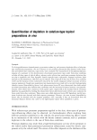
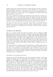
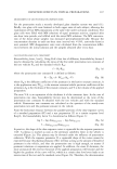
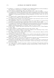
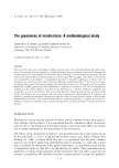
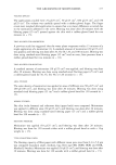
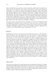
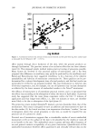
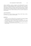
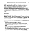
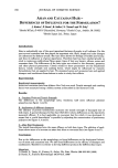
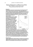
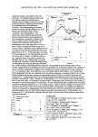
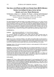
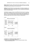
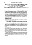
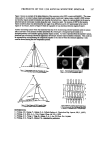
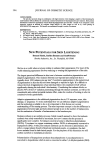
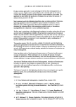
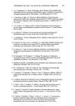
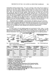
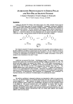
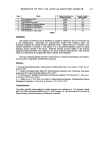
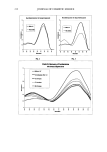
PREPRINTS OF THE 1998 ANNUAL SCIENTIFIC SEMINAR 191 conduction through a semi-infinite solid, were performed. The calculated data have shown that near-uniform temperature distributions are ,reached in the hair samples within a few seconds of thermal exposure, suggesting that continuous and intermittent modes of treatment are equivalent. The resulting damage to the fibers has been investigated and quantified by the use of fluorescence spectrophotometry, Hunter colofimetry, and combing analysis. The fluorescence analysis analysis has shown that thermal treatment results in a decomposition of hair chromophores, specifically tryptophan (Trp) and its oxidation products (kynurenines). The calculated first-order rate coefficients of Trp decomposition were in the range from 0.03 to 0.12 (rain -I) with an estimated activation energy of 6.6 kcal/mol. Figure 1 illustrates the decomposition of Trp as a function of the thermal treatment time for various types of hair at 164 øC. At this temperature, the extent of Trp decomposition is very high reaching nearly 80% conversion after 30 minutes. In addition to this, we have employed Hunter colorimetry to quantify the thermally induced color changes in hair, such as an increase in the yellowhess of white and Piedmont hair, or simultaneous yellowing and darkening of bleached it 1o o o Hair Thermal Damage Figure 2 ,m•. ] Reg•n .o, f Curling iron , 4O 0O stun !Oe 120 140 toe Di ce (ram) 2O • PVP•DMAPA Ac•ate* CopoJym or ß •uatemlmu 70 x unb•eated 7o. aeoo 4 0 8( !0 '$2 ?line rain) Figure 3 hair. Surface damage, resulting from thermal treatment, was quantified by using combing analysis, which revealed a gradual increase in combing forces that were measured in the tress section exposed to the curling iron. Figure 2 provides a representative example of the combing force increases resulting from thermal exposure at 152 øC for light brown hair. Force difference curves were determined by subtracting the curve obtained after thermal degradation from the curve obtained prior to any thermal treatments. A possible method for preventing thermal damage to hair has been tested by pre-treating fibers with PVP/DMAPA Acrylates Copolymer and Quatemium 70. The decomposition of Trp in fibers pretreated with the indicated actives, followed by thermal exposure, was monitored. Figure 3 presents the Trp degradation at 152 øC as a function of the thermal treatment time for light brown hair. As indicated by the data in Figure 3, depositing a layer of surfactant on the surface of hair appears to provide some degree of Trp protection. We also monitored the surface modifications of the fibers, resulting from thermal treatment at 152 øC, by combing analysis. Figure 4 pertrays combing work difference as a function of time for hair treated with the indicated actives and for untreated hair. The combing work difference values were determined by integrating the area of the combing curve where thermal treatment was administered and subtracting this value from the similarly obtained value prior to thermal exposure. Similar studies were conducted at 132 øC and provided comparable data with respect to the possible thermal protection properties of the tested compounds. x tMiJ. elfod x Figure 4 o o lO 12 Time (rain) References [1] P. Milczarek, M. ZieLinski, and M. Garcia, Colloid Polym. Sci., 270, 1106 (1992). [2] R. Crawford, C. Robbins, and K. Chesney, J. $oc. Cosmet. Chem., 32, 27 (1981). [3] L. Rebenfeld, H. Weigmann, and C. Dansizer, o r. $oc. Cosmet. Chem., 17, 525 (1966). [4] W. Humphties, D. Miller, and R. Wildnauer, or. Soc. Cosmet. Chem., 23, 359 (1972). [5] R. Amaud, G. Perbet, A. DeFlandre, and G. Lang, Int. J. Cosmet. Sci., 6, 71 (1984). [6] C. Pande and J. Jachowicz, or. $oc. Cosmet. Chem., 44, 109 (1993).

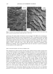
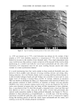
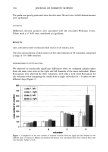
192 JOURNAL OF COSMETIC SCIENCE THE ISOLATED PERFUSED BOVINE UDDER SKIN (BUS) MODEL: SURFACTANTS/SKIN INTERACTIONS AFTER SHORT AND LONG TERM EXPOSURE W. Pitterman •, J. Kahre •, H. - U. Kriichter 2, Th. Fi•rster 1, L. Kintrup • and M. Kietzmann 3 •Henkel KGaA, D-40191 DiisseldorjS, Germany :Henkel Corp., Ambler, PA 19002 31nstitute for Pharmacology, Pharmacy and Toxicology, Vet. School Hannover, D-30559, Hannover, Germany INTRODUCTION: The isolated perfused bovine udder skin model (1) was originally introduced as an in vitro model to study the percutaneous absorption. Under in vitro conditions barrier properties and metabolization comparable to living skin are maintained in perfused skin models. Due to this viable status additional information concerning time- dependent skin irritation can be obtained in punched skin biopsies representing epidermal and derreal layers. The model gives the opportunity to distinguish cytotoxicity (modified methyl tetrazolium assay) (2) from irritancy (prostaglandin E2-tissue concentration). The period of exposure (0.5 hour up to 5.0 hours) as well as the type of application (open / occluded Finn Chamber R) is variable within the perfusion time of approx. 8 hours. EXPERIMENTAL: Surfactants: Alkyl polyglycoside [APG] (pH.: 5.5 3 % and 10 % active substance) Sodium Lauryl Sulfate [SLS] (pH.: 5.5 3 % and l0 % active substance) The unpreserved surfactants were handled as frozen samples during the logistic procedures before the application. The surfactants were applied occlusively using a chamber (D=18 mm 500pl / Finn Chamber ©) under adhesive tape. Model: The test set-up is extensively described (1). The viability of the perfused udder was demonstrated by a nearly unchanged glucose consumption, an initially decreasing and thereafter unchanged lactate production and an unchanged dehydrogenase activity in the perfusate after it has flowed through the vascular system. It may be assumed that no significant edema developed within 6 hours since the skin fold thickness was constant. Morphology: Samples of the treated and untreated skin underwent a routine preparation for H&E histology and transmission electron microscopy. Biochemistry: Cytotoxicity: The MTT-assay indicates the level of impaired mitochondria in the cells of the epidermal layers and derreal tissue. The modified MTT (methyl tetrazolium salt dye conversion, pg Formazan / pg DNA) is used (Maag, 1993). Irritancy: The results of the assessment of the prostaglandin E2-concentration (ng/pg DNA) in the epidermal and derreal layers presents the level of a mediator substance responsible for erythema and edema formation in the case of clinically evident skin irritation (MaaB, 1993). For both assays the whole skin biopsy (D=6mm, approx. 4 mm depth) is used. Due to the different cell concentrations in the epidermal and derreal layers, the tissue preparations were adjusted to the comparable DNA content before being analyzed. For the statistical analysis the t-test was used. CONCLUSION: Cytotoxicity (MTE, Figure/):After an occlusive exposure period of 1 hour the application of APG 3 % and 10 % AS does not induce a statistically significant cytotoxic potential compared to the untreated skin area whereas the application of SLS 3 % and 10 % resulted in a statistically significant difference (p 0.05). After 1 hour of exposure the cytotoxicity may be predominantely influenced by physicochemical processes which in the case of SLS was much more pronounced than when applying APG. After an exposure period of five hours the cytotoxic potentia, l shows a comparable level (p 0.05). This demonstrates that the exaggerated test conditions allow a skin penetrating effects of the surfactants which is much less pronounced in the case of APG than when using SLS. APG 10 % AS has about the same potential as APG 3 %or SLS 3 % AS. SLS 10 % AS after a period of exposure of 1 hour and 5 hours impairs about 30 % and 50 % resp. of the epidermal and dermal cells.
Purchased for the exclusive use of nofirst nolast (unknown)
From: SCC Media Library & Resource Center (library.scconline.org)