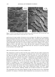
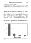
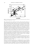
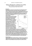
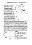
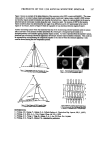
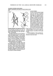
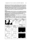
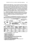
PREPRINTS OF THE 1998 ANNUAL SCIENTIFIC SEMINAR 199 TRANSIENT NETWORK COPOLYMERS- SURFACE PROPERTIES AND ASPECTS OF SKIN INTERACTIONS Cont figure 1 hydrogels described in this paper are non-tacky, leave uniformly thin films on the skin. are reversibly shear-thinned (thixotropic), tolerate elec- trolytes and pH extremes very well, and confer a 'silky', low friction feel. The ability of the transient network hydrogels to deform and reform accounts for several of their unique and desirable properties. Lack of tackmess in these systems is a result of the structural flecibil- ity (non-covalent bonding) of the hydrogel's matrix, as is the ease of spreading. The thixotropic character of the hydrogel matrix, likewise, enhances uniformity of film thickness and the resultant distribution of active ingredients. The topic polymers of this paper orient so that the hydrophilic portions am in contact with the skin and the hydrophobic portions form a protective layer above. This not only accounts for the pleas- ant after-feel, but also produces a protective hamer that reduces TEWL. The segmented hydrophilic- hydmphobic segmentation of these polymers also gives them the ability to cross the oil-water inter- face and assist in emulsion stabilization. and non-tacky properties, cast uni- form, continuotis films and assist in the stabilization of emulsions typical of skin-care formulations. The pre- sentation of hydrophobic moleties to the environment confers a smooth feel to the skin and provides a protec- tive barrier that reduces TEWL. Unlike hydrogels derived from covalently crosslinked polymers, the subject hydrogels are unusually resistant to electrolyte level and pH extremes. All of these properties. as well as numerous oth- ers. are very. beneficial to meet the emerging demands of the skin-care market. CONCLUSIONS Transient network hydrogels formed from non-covalently crosslinked polymers offer many significant advantages over traditional hydro- gels. Our work, based on the devel- opment of controlled base-catalyzed polyacrylonitrile polymers, has shown that hydrogels derived from these polymers display thixotropic

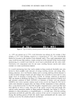
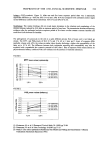
200 JOURNAL OF COSMETIC SCIENCE The Clinical and Laboratory Assessment of Skin Whitening Walter Smith, Ph.D. Dermac Laboratory, Stamford, CT and Scott Norton, Ph.D., University of Texas, Denton Skin whitening can be evaluated with a variety of in vitro and in vivo test methods with duel goals of optimizing product performance and providing substantiation for product claims. Test tube assays monitoring the tyrosinase dependent conversion of tyrosine to L-Dopa have been useful in comparing the activity of tyrosinase inhibitors. Our tests indicate that kojic acid, ascorbyl magnesium phosphate, Mulberry extract, and numerous herbal extracts are effective enzyme inhibitors. This in vitro activity however does not always translate into clinical effectiveness. In a different test system we have evaluated the inhibition of delayed hyper-pigmentation resulting from complete removal of the stratum corneum via tape stripping. This in vivo test has confirmed activity of kojic acid, and some Mulburry extracts, however vehicle differences could markedly influence activity, and correlation with the in vitro assay was poor. Finally we have evaluated the skin whitening effects of several cosmetic prototypes over a three to four month period. Clinical grading, photography, and the Minolta Meter have proved useful in assessing skin color. Good correlation is observed with the three methods, with clinical grading being most sensitive. We have observed that herbal extracts, hydroquinone, and combinations of AHA's can induce measurable and consumer relevant skin lightening after two to three months of product use. In most cases a general skin lightening is observed without preferential lightening of "age spots". AN IN VITRO METHOD FOR SCREENING WHITENING AGENTS Gopa Majmudar, Ph.D., George Jacob, Yolanda Laboy and Lou Fisher, Ph.D. Mary Kay HoMing Corporation, Dallas, TX 75247 INTRODUCTION: The development of successful whitening skin care products depends on the use effective whitening or depigmenting ingredients that reduce melanin production in melanocytes. We utilized a three dimensional human skin model, Melanoderm, to screen whitening agents before testing the product on humans. Melanoderm is a living skin equivalent in vitro model of the human epidermis consists of well differentiated human keratinocytes and melanocytes. Biochemical, histological and ultrastructural properties of Melanoderm are sim- ilar to human epidermis. The cross section of the Melanoderm shows the presence of stratum corneum, keratinocytes and dendritic melanocytes localized in the basal cell layer. Melanocytes stain positive when exposed to L-dihydroxyphenyl alanine (L-DOPA), a precursor of melanin. A relative activity of whitening agents such as kojic acid, lactic acid and magnesium ascorbyl phosphate (MAP) was measured on Melanoderm. Total kojic acid in the product was measured by high performance liquid chromatography (HPLC). A combination of in vitro test on human skin equivalent culture and HPLC method is useful to evaluate the efficacy, stability and toxicity of whitening ingredients and products before clinical testing. Finally, the product was tested on human subjects to obtain a better correlation with in vitro test results. MATERIALS AND METHODS: In Vitro Testing: Melanoderm was purchased from Mat Tek Co, Ashland, MA. Kojic acid, lactic acid and MAP in aqueous or anhydrous base and the base alone were applied to Melanoderm and incubated for two to three days. At the end of incubation, the cream was removed. Melanoderm was washed in phosphate buffer saline (PBS) and incubated in 0.1% L-DOPA for one hour at room temperature. After one hour Melanoderm was placed in 0.1% fresh L-DOPA and incubated 4 to 16 hours. Optical density was measured at 490rim using a microplate reader. HPLC: Kojic acid assay was performed using a HP laser jet system with a four plus integrator. A C s, ODS -5, 150 X 4.5 mm column (Metachem//0297) and a mobile phase composed of water acetonitrile (20:80 v/v) were used. The wavelength of detection was 254nm. Samples were diluted with water:acetonitrile (50:50 v/v).
Purchased for the exclusive use of nofirst nolast (unknown)
From: SCC Media Library & Resource Center (library.scconline.org)