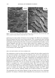
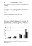
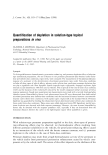
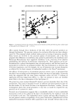
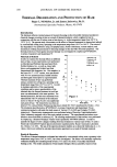
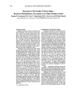
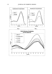
PREPRINTS OF THE 1998 ANNUAL SCIENTIFIC SEMINAR 203 hyperpigmentation and to make judgments based on pigmentation differences in the upper cheek and eye area only. The judge indicated which of the two images had less noticeable hyperpigmented spots and rated the magnitude of the difference between images on a 1-4 scale. The magnitude rating was assigned a positive (+) or negative (-) sign depending upon whether (+) or not (-) the judge indicated the post-treatment image had less noticeable spots. Thus, positive values of the magnitude rating indicate spot reduction efficacy and values of zero or less indicate no efficacy. Statistical Analysis: The difference between the test treatments' (SLM and E) mean change from baseline and their respective controls' (NM and V) mean change from baseline for L*, a*, and b* values and hyperpigmented spot area were compared using a paired T-test (significance set at p0.05). The mean VPS grades for each treatment and control pair were also compared using a paired T-test. A non-parametric statistic analysis (McNemar's test) was used to compare the test products for the number of 'AFTER' images selected as improved in the VPS analysis. RESULTS: Reproducibility and Accuracy: The within subject COVs for L*, a*, and b* value measurements in the cheek and eye area were 0.6%, 2.1% and 1.8•/• respectively. The within subject COV for the hype•igmented spot area measurement in the cheek and eye area was 0.5%. The imaging system accuracy for measuring changes in hyperpigmented spot area was determined to be less than +/-5% when measurements were made in the region of the cheek and eye area. All image analysis measurements of live subjects were confined to the area of the cheek and around the eye. Subiect Accountability: Of the 120 subjects enrolled, 104 completed the study (118 completed through 3 months). Basal Skin Color Tone: There was no significant difference between treatments for L*, a* or b* value change from baseline (see Table I for L*-valuc change from baseline). Hyperpigmented Spot Area: Both test treatments (SLM and E) showed significant reduction in hypcrpigmented spot area vs. their respective controls at all time points (Figure 2). Visual Perception System {VPS}: The mean VPS grades for the test treatments (SLM & E) were significantly greater than their respective controls at all time points (Figure 3). Judges more often selected the AFTER image (vs. the BEFORE image) for both SLM & E vs. their respective controls (Table II). Figta'e 4 shows hyperpigmentation improvement on one subject treated with E on one side of her face at baseline and 6 months (image overlays are also shown). FIGURE I TABLE I TABLE II Asian Shaped Mannequin Heads L*-value Change From Baseline VPS % AFTER Images Selected Months Months Treatment I 3_ 6 SLM -0.27 0.78 0.04 NM -0.26 0.96 -0.20 E -0.27 0.61 0.22 V -0.19 0.55 0.07 Treatment ! 3_ f SLM 74* 60* 77* NM 45 37 56 E 60* 43* 62* V 40 30 43 *Sig. diff. from control (p0.025), McNemar's Test FIGURE 2 FIGURE 3 FIGURE 4 Hyperpigmented Spot Area VPS Mean Grades Subject Treated With Product E Baseline image (left) with overlay (right) SIg. diff. frown NM (pO.001) ' • ]T•g. diff. frown NI4 (pO.001) i Monks of T•a•ent Monks of T•a•ent 6-mon• image (le•) with overlay (right) Monks of Tm•ent Mon• of T•a•ent

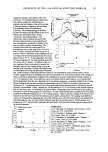
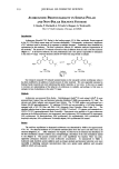
204 JOURNAL OF COSMETIC SCIENCE CONCLUSIONS: The split-face protocol design in combination with high resolution video imaging is capable of discriminating the small treatment effects afforded by commercial cosmetic formulations for hyperpigmented spot reduction. Both product formulations tested here significantly reduced facial hyperpigmented spot area aRer 1 month of use compared to their respective controls as assessed by computer image analysis of video images as well as by visual grading of BEFORE/AFTER image pairs. This benefit was maintained through 6 months of treatment. REFERENCES: 1. Kang S. and Sober AJ., "Disturbances of Melanin Pigmentation", in Dermatology, Moschella and Hurley, Ed., 3 rd Ed., Vol. 2, 1442-1474 (1992). 2. M. Jimbow and K. Jimbow, Pigmentmy Disorders in Oriental Skin, Clinics in Dermatology, 7, 11-27 (1989). 3. Caron D. et al., Split -face Comparison of Adapalene 0.1% Gel and Tretinoin 0.025% Gel in Ache Patients. JAm AcadDermato136 (6 Pt 2) S110-112 (1997). NEW POTENTIALS FOR SKIN LIGHTENING Durant Scholz, Suelien Bennett and Geoff Brooks Brooks Industries, Inc., So. Plainfield, NJ 07090 We live in a world where everyone wishes to enhance their appearances. For most of the world enhancing appearance involves reducing or evening the pigmentation of the skin. The largest perceived difference in skin tone is between constitutive pigmentation and adaptive pigmentation. This variation between sun-exposed and unexposed skin is typically about 1000 melanocytes per mm 2. Consfitutive pigmentation is the natural level of pigmentation in skin that has not been damaged or exposed to light. It is difficult to reduce the level of pigmentation of an individual below the constitutive level without significantly altering the individual's biochemistry. Considering that melanin blocks or absorbs 90% of the UV radiation penetrating through the stratum corneum, as well as the considerable anti-oxidant potential melanin offers, make the wisdom of looking to alter its constitutive levels questionable. Adaptive pigmentation is the additional pigmentation due to UV exposure, acne, skin damage, or pregnancy. To truly understand how we can influence adaptive pigmentation and the technology available to do so it is important to first discuss our current understanding of the tanning process. The most important concept is that the difference in pigmentation between light and dark skinned people is not in the number of melanocytes but the mechanism of transfer of melanosomes to the keratinocytes. Melanin synthesis is an oxidative process. Initial research seemed to show that melanin synthesis was solely controlled by tyrosinase, but now it seems that the process is significantly more complex. Tyrosinase converts tyrosine to dihydroxy phenylalanine (DOPA). Tyrosinase is a copper containing oxidase. DOPA is subsequently converted to dopaquinone (DQ). At this point melanin synthesis can be directed to pheomelanin, the yellow-orange melanin found in blond and red-haired people, or eumelanin, the dark brown melanin found in dark-haired people. For the purposes of this discussion we will confine ourselves to eumelanin production. Polymerization of dopaquinone produces leucodopachrome. Leucodopachrome is converted to dopachrome which in turn is converted to either 5,6 dihydroxyindole (DHI)
Purchased for the exclusive use of nofirst nolast (unknown)
From: SCC Media Library & Resource Center (library.scconline.org)