PREPRINTS OF THE 1998 ANNUAL SCIENTIFIC SEMINAR 207 A. K. Chakraborth, S. J. Ofiow, JL Bolognia, and JL Pawelek, Structural/functional relationships between internal and external MSH receptors: modulation of expression in Cloudman melanoma cells by UVB radiation, J Cell Physiology 147 1 (1991) C. Bertolotto, K. Bille, J.P. Ortonne, R. Balloff, Regulation oftyrosinase gene expression by cAMP in B 16 melanoma cells involves two CATGTG motifs surrounding the TATA box: implication of the micropthalmia gene product. J. Cell Biol. 134: 747- 755 (1996) P. R. Gordon, C. P. Mansur, and B. A. Gilchrest, Regulation of human melanocyte growth, density, and melanization by kerafinocyte-derived factors., J. Invest Dermatol 92 565 (1989) 8. G. Imokawa, Y. Mishima, Active tyrosinase site in melanin synthesizing cells: Biochemical analysis of coated vesicles, Jap J Dermatol 88 913 (1978) 9. J. Vachtenheim, J. Duchon, Melanogenic factors: regulation ofgene expression. Sb Lek 97:41-47 (1996) 10. I. Suzuki, R. D. Cone, S. Im, J. Nordlund, Z. A. Abdel-Malek, Binding ofmelanotropic hormones to the melanocortin receptor MC 1R on human melanocytes stimulates proliferation and melanogenesis. Endocrinology 137:1627-1633 (1996) 11. G. Imokawa, Y. Yada, M. Kimura, Signaling mechanisms of endothelial-induced mitogenesis and melanogenesis in human melanocytes. Biochem. J. 314:305-312 (1996) 12. K. Kameyama, P.M. Montague, V. J. Hearing, The expression ofmelanocyte- stimulating hormone receptors correlates with mammalian pigmentation and can be modulated by intefferons. J Cell Physiol 137:35-44 (1988) 13. Signaling mechanisms of endothelin-induced mitogenesis and melanogenesis in human melanocytes. Biochem J. 314:305-312 (1996) 14. B. A. Gilchrest, H. Y. Park, M. S. Eller, M. Yaar, Mechanism of ultraviolet light- induced pigmentation. Photochem Photobiol. 63: 1-10 (1996) 15. T. Nishimura, T. Komentani, S. Okada, N. Ueno, T. Yamamoto, Inhibitory effects of hydroquinone-alpha-glucoside on melanin synthesis. Yakugaku Zasshi 115:626-632 (1995) 16. Y. Mishima, Molecular and biological control ofmelanogenesis through tyro sine genes and intrinsic and extrinsic regulatory factors. Pigment Cell Res 7495:376-387 17. S. Ando, O. Ando, Y. Suemoto, Y. Mishima, Tyrosinase gene transcription and its control by melanogenic inhibitors. J. Invest Dermatol 100: 150S-155S (1993) 18. Y. Tomita, W. Torinuki, H. Tagami, Stimulation of human melanocyte by vitamin possible mediates skin pigmentation at%r sun exposure. J. Invest Dermatol. 90:882 (1988) 19. P. Valverde, E. Benedito, F. Solano, S. Oaknin, J. A. Lozano, J. C. Garcia-Borron, Melatonin antagonizes alpha-melanocyte-stimulating hormone enhancement of melanogenesis in mouse melanoma cells by blocking the hormone-induced accumulation of the c locus tyrosinase. Eur J Biochem 232:257-263 (1995)

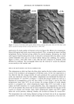
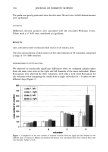
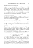
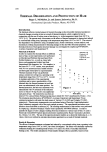
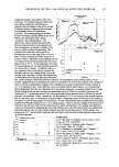
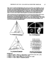
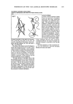
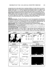
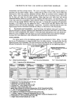
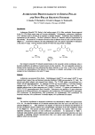
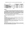
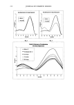
208 JOURNAL OF COSMETIC SCIENCE A COMPARISON OF SKIN LIGHTENING AGENTS Evelyn G. Su, Ph.D. Sino Lion (USA) Ltd., New York, NY 10048 INTRODUCTION: Skin lightening products have become increasingly popular in the Asian-pacific countries as well as in the African and South American countries. Europe and America have also seen increased interests in skin lightening agents. For Asian and Black skin, the main purpose for skin lightening products is to lighten or whiten the skin as well as to even-tone and brighten the skin while for Caucasian white skin the focus is on even-toning and brightening the skin. For all skin types, the skin lightening agents can be used to treat pigmentation disorders such as freckles, pregnancy masks and age spots. The purpose of this paper is to compare the various aspects of the four popular skin lightening agents, i.e., kojic acid, kojic dipalmitate, arbutin and magnesium ascorbyl phosphate (MAP), as well as hydroquinone. SKIN COLOR AND MELANIN: Skin color is mainly determined by the amount of melanin present in the skin. Melanin is synthesized in melanocytes which are normally found in the epidermal basal layer. Within the melanocytes melanin is bound to a protein matrix to form melanosomes. In the melanosomes, tyrosinase converts tyrosine to eumelanin or pheomelanin. Fig. 1 illustrates the pathways of melanin biosynthesis in the melanocytes. By blocking at the various points of the pathways, skin lightening agents can inhibit or even reverse melanin biosynthesis, and are thus useful in whitening or lightening the human skin. Skin lightening agents can also be used to treat local hyperpigmention or spots which are caused by a local increase in melanin synthesis or uneven distribution. SKIN LIGHTENING AGENTS: A. Mechanism of Action: Arbutin, kojic acid, kojic dipalmitate, MAP are all tyrosinase inhibitors. Arbutin reportedly works by competing with DOPA at its receptor site on tyrosinase, • while kojic acid inactivates tyrosinase by chelating with its vital copper ion and suppressing the tautomerization from dopachrome to DHICA) L-ascorbic acid and its derivatives, which include MAP, are believed to act as reducing agents on melanin intermediates, thus blocking the oxidation chain reaction at various points from tyrosine/DOPA to melanin) Kojic dipalmitate is a tyrosinase inhibitor, but the exact mechanism of action is unclear. Hydroquinone is also a tyrosinase inhibitor. n' s In addition, hydroquinone has cytotoxic effect on melanocytes, which causes high toxicity to the skin. B. Ease of Formulation and Stability: Kojic Acid, arbutin and MAP are all water-soluble, and can be easily incorporated into the formulation. On the other hand, kojic dipalmitate is oil-soluble, and can be easily melted into oil phase at 75-85øC, followed with immediate emulsification. However, kojic acid, hydroquinone and arbutin have color stability problems, which impose various difficulties on formulation. Kojic acid usually tums yellowish brown with time in the finished product. The reasons for this instability are mainly two-fold: a) kojic acid chelates with many metal ions, especially for iron (Fe3+), to produce colored complex (yellow color for iron) b) kojic acid can oxidize slowly in contact with air, and this process accelerates at high temperature. Hydroquinone solution becomes brown on exposure to air as a result of oxidation and must be stabilized. Arbutin has by far higher stability than hydroquinone, 6 but still has potential instability in formulation. MAP is stable in formulation. Kojic dipalmitate is stable to heat, light, pH and oxidation. Compared with kojic acid, kojic dipalmitate neither chelate with metal ions and nor oxidize in air. As a result, kojic dipalmitate has excellent stability in formulation. C. Safety and Efficacy: Kojic acid is non-toxic and has minimal irritation. 7 Kojic acid has been used in Japan since 1988 and has extended to other parts of the world. The human patch test on kojic dipalmitate showed that it is completely non-irritating. 8 There are no known safety issues associated with the use of kojic dipalmitate. Hydroquinone can be irritating and cause redness and burning. More importantly, it has been shown to cause exogenous ochronosis, 9' •0 which has resulted in a ban on its use in South Africa, Thailand and other countries. Arbutin has far higher safety than hydroquinone, has no irritation and virtually no sensitization) • MAP is a vitamin C derivative, has an excellent safety record. Figure 2 shows the comparison of tyrosinase inhibitory effects among kojic acid, hydroquinone and the blank (water). The y-axis is absorbency at 475 nm which measures the coloration of various melanin
Purchased for the exclusive use of nofirst nolast (unknown)
From: SCC Media Library & Resource Center (library.scconline.org)