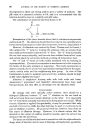
38 JOURNAL OF THE SOCIETY OF COSMETIC CHEMISTS no uptake of labelled tyrosine. The ability to oxidise tyrosine is thus found in both melanic and pheomelanic follicles, melanic animals showing a greater activity than pheomelanic. It is quite clear at this time that black and brown. melanin appear to be closely related chemically, and genetical evidence indicates that their modes of formation are closely similar. Pheomelanin differs chemically from melanin, and genetical evidence indicates a very distinct mode of formation for the two pigments. Tyrosinase is involved in the formation of both melanin and pheomelanin, and tyrosine can be considered to be the precursor of melanin. While tyrosine will act as a substrate for ph•.omelanic hair follicles in vitro, the pigment formed is abnormal, and there is little or no indication that tyrosine is the natural chromogert of pheomelanin the oxidation of tyrosineby pheomelanic follicles may be involved only indirectly in pigment formation, and tyrosine (or its oxidation products) may not be the pigment precursor. The •ctivity of the genes for pheomelanin production in the guinea pig or the mouse "turn on" the production of pheomelanin in a very definite way no intermediates between melanin and pheomelanin appear to be formed. This clear-cut action of the genes presupposes a switch- mechanism, probably involving one enzymic step. The possible dual role of tyrosine and tryptophane intermediates in this switch-mechanism is suggested by the investigations of Butenandt, who showed that the formation of the red-yellow pigment, xanthomatin, depended on the conversion of dopa to dopa quinone in the presence of tyrosinase, and the non-enzymic oxidation of 3-hydroxykynurenin to xanthomatin by the dopa quinone which was reduced back to dopa. In some histochemical experiments using red human hair balbs and hair follicles of intense yellow, e/e, guinea pigs, we have demonstrated that melanin formation is absent in pheomelanic human and guinea pig hair follicles following incubation in dopa and 3-hydroxykynurenin in a molar ratio of 1: 4 for 20 hours. If the ratio of these substrates is 4:1 (dopa: 3-hydroxykynurenin) no black pigment is formed in four hours, but after 20 hours (all the 3-hydroxykynurenin having been oxidised) black pigment is found to have been deposited. These results provide necessary but not sufficient evidence for an explanation of pheomelanin formation as the result of the oxidation of an o-aminophenol by dopa quinone produced by the oxidation action of tyrosinase on dopa. It is compatible with the observation that pheomelanic hair follicles contain tyrosinase, and it would explain the pigmentary switch-mechanism leading either to melanin or pheomelanin, this being the result of the absence or presence of o-amino- phenol. The critical enzyme operating the switch could be one bringing about hydroxylation of an aromatic amine. The yellowish-brown pigments formed by the oxidation condensation of o-aminophenols are soluble, as is pheomelanin, in dilute alkali. Although no systematic search for trypto-

ABSTRACTS OF PAPERS ON THE BIOLOGY OF HAIR GROWTH 39 phane intermediates has been made in hair, Rebell has recently isolated kynurenin from the yellowish hair of albino rats. In addition, we have observed that pheomelanic hair when illuminated with wave-lengths of 3,600 A. is fluorescent, and emits light varying from a dull orange to bright yellow. Yellow hair, the pheomelanic banding of agouti hair of rn•ice, guinea pigs and golden hamsters and human red hair show this effect. The fluores- cence of the mixture of 3-hydroxykynurenin and dopa in which hair follicles had been incubated was a dark orange colour resembling that of an alkaline extract of pheomelanic hair. "THE BEHAVIOUR OF PIGMENT CELLS AND EPITHELIAL CELLS IN THE HAIR FOLLICLE" HERMAN B. CHASE Biology Dept., Brown University, Providence 12, Rhode Island. In the germ region of the regenerating hair follicle there are two centres of growth, one around the dermal papilla and giving rise to the bulb, the other producing the elongated lower external sheath. The first portion of the inner sheath forms from the early bulb and more is added as the bulb descends. The cells of the matrix, the lower bulb, are indeterminate in that they can be mechanically displaced and still give rise to the three layers of the inner sheath and the three regions of the hair shaft. Melanocyte stem cells reside in the upper bulb which is derived from the germ region capping the dermal papilla. The mature melanocyte has short dendrites, the cell body projects into the cavity of the dermal papilla and it delivers granules to be engulfed by the recipient hair cells. The club and surrounding capsule are formed at catagen from cells of the bulb matrix, but only after the dermal papilla has become partially rounded. "THE ELECTRON MICROSCOPY OF THE MELANOCYTE" N. A. BARNICOT University College, University of London, London, W.C.1, England, and IV[. S. C. BIRBECK Chester Beatty Research Institute, Royal Marsden Hospital, London, S.W. 3, England. The examination of melanocytes from human hair bulbs with the electron microscope has allowed the mechanism of pigment granule formation to be studied. A.region near the nucleus of the melanocyte, corresponding to the Golgi region, contains numerous small vesicles these consist of an outer membrane with several concentric inner membranes. Melanin is deposited upon the vesicles, to form the completed pigment granules. These are then transferred to the cortical cells of the hair. Examination of albino hairs has shown that the complex membrane structures are synthesised by the melanocyte, but
Purchased for the exclusive use of nofirst nolast (unknown)
From: SCC Media Library & Resource Center (library.scconline.org)