j. Soc. Cosmet. Chem., 36, 349-354 (September/October 1985) X-ray diffraction study of human stratum corneum STIG E. FRIBERG and DAVID W. OSBORNE, Chemistry Department UMR, Rolla, MO 65401, and THOMAS L. TOMBRIDGE, Pathology Department, St. John's Regional Health Center, Springfield, MO 65804-2263. Received December 17, 1984. Synopsis A study of human thin skin stratum corneum was made using small-angle x-ray diffraction. This study elucidated how a new type of skin softener, 2-(alkoyloxy)-l-[(alkoyloxy)methyl]-ethyl-7-(4 heptyl-5,6- dicarboxy-2-cycloheoxene-l-yl ) heptanoate (G2), interacts with the stratum comeurn. A broad diffraction peak from 50-80 A, assigned to the lipid content of the stratum comeurn, was characteristic of untreated normal stratum corneurn. Washing of the specimen with a 0.1% soap solution removed the 50-80 • peak, while treatment with G2 caused the occurrence of a diffuse diffraction peak from 30-45 • for both unwashed and prewashed stratum comeurn. Treatment with a triglyceride oil gave no change in the diffraction pattern. It appears obvious that G2 has a penetrative action on stratum comeurn that is not present for unmodified triglycerides. INTRODUCTION Small-angle x-ray diffraction studies to determine the structure of human stratum corneum were first carried out by Swanbeck in the late 1950s (1,2). This landmark work provided a model that depicted the stratum corneum as being composed of 250 fk protein bundles surrounded by 80 fk thick lipid layers. Since this study, small-angle x-rays have been used by other investigators to examine the stratum corneum and the epidermal lipid it contains (3,4). These studies have mainly used stratum corneum samples obtained from thick skin. However, as has been demonstrated (5), the barrier function and physical properties of thick skin stratum corneum are significantly different from those of thin skin stratum corneum. Since thin skin covers most of the body, especially those parts targeted by ß cosmetics, investigation of the properties of its stratum corneum are of obvious signif- icance. In addition, it is reasonable that information gained from investigations on thick skin may not be totally applicable to the thin skin counterpart, since thin skin provides a much greater barrier function through its different structure. Earlier diffraction studies concentrated on structural elucidation of the stratum corneum for both healthy skin and diagnosed dermatological conditions. However, there is no reported use of low-angle x-ray diffraction to study the effects of external treatments on the stratum corneum. Even the effect of washing upon the epidermal lipids has not been studied, despite the use of thorough washing with Ivory © soap by sebum donors two hours prior to collection (6). It was such experiments by Downing and co-workers 349

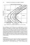
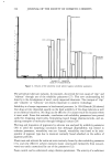
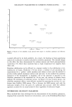
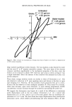
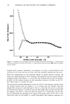
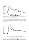
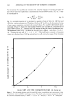
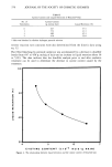
350 JOURNAL OF THE SOCIETY OF COSMETIC CHEMISTS (6) that produced much of the current knowledge concerning the composition of human sebum. Xerosis, the condition commonly referred to as dry skin, is a condition that is considered almost universal among the aged (7). We found the potential for a direct investigation of a cosmetic treatment on stratum corneum of great appeal and used small-angle x- ray diffraction techniques to investigate the effects of washing, treatment with G2, and ether extraction upon the stratum corneum of human thin skin samples. EXPERIMENTAL Fresh full thickness samples of human skin were obtained from radical mastectomy cases and prepared by a method described by Lampe et al. (8). The skin samples were from normal skin well away from the tumor site. After removal of the subcutaneous fat with a #22 blade, each sample was placed dermis side down on filter paper soaked with 0.5 % trypsin in phosphate-buffered saline (PBS). The samples were then incubated while refrigerated for 24 hours prior to removal of the stratum corneum. Sheets of stratum corneum were then thoroughly rinsed with PBS and frozen on filter paper after being blotted dry. Immediately prior to x-ray analysis, the samples were thawed and vortexed in PBS to remove any granular cells or filter paper residue. The samples, taken from the same skin specimen, were then prepared for low-angle x- ray measurements by one or more of the following techniques. "Washed" samples were vortexed in a 0.1% (by weight) Ivory © soap solution for ten minutes. Samples "treated with G2" were spread on filter paper soaked with purified G2 and allowed to remain in contact with G2 for one to thirteen hours. "Extracted" samples were vortexed in ethyl ether for 30 minutes. The prepared stratum corneum sheets were then rolled and placed in a 0.7-mm glass capillary tube and examined by small-angle x-ray diffraction. The untreated stratum corneum band was checked by multiple measurements of skin specimens from three different subjects, while the treated, washed, and extracted sam- ples were determined by duplicate measurements from the same skin specimen. Only slight differences in intensity were encountered between spectra of the multiple runs. G2 was purified as described previously (9). Trypsin (98%) and dry phosphate-buffered saline (diagnostic) were obtained from Sigma Chemical. Absolute ethyl ether (Aldrich, Reagent) was used for extraction. All glassware was thoroughly cleaned and ultimately rinsed with ether to eliminate the possibility of external lipid contamination. Small-angle x-ray diffraction spectra were collected for seven hours using a Kiessig low- angle camera from Richard Siefert. Ni-filtered Cu radiation was used and the reflections determined by a Tennelec position-sensitive detection system (Model PSD-1100). RESULTS Normal stratum corneum gave a characteristic broad diffraction peak of moderate in- tensity in the range of 50-80 • (Figure lo). No other bands were distinguishable over the coml•lete available range of 20- 160 A for collection times of twelve hours. This 50-80 A band was removed both by washing and by extraction with ether as seen in Figure 2.
Purchased for the exclusive use of nofirst nolast (unknown)
From: SCC Media Library & Resource Center (library.scconline.org)