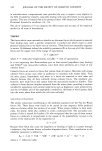
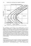
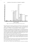
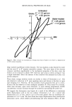
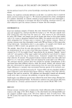
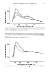
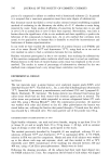
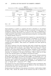
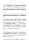
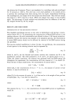
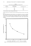
STRATUM CORNEUM X-RAY DIFFRACTION 351 600 400 oøO o o o o o o o o Oo o o o o o • [] Uoo D o a a a oo_oo o o o o o ß ß ß ! sb ' eo INTERLAYER SPACING (•) Figure l. Effect of G2 on x-ray diffraction spectrum of washed skin. O, untreated normal stratum corneum [U, washed normal stratum corneum ', washed normal stratum corneum treated with G2 for one hour O, region of complete spectral overlap. Treatment with G2 produced a weak diffraction from 30-45 fk. The intensity of this diffraction increased slightly as the treatment duration increased from one hour to thirteen hours (Figure 3), while the intensity of the 50-80 /k band significantly de- creased for the same change in treatment duration. Treatment of the stratum corneum with soy oil for one hour produced a narrower, slightly shifted diffraction peak from 60-80 fk. This spectrum is compared to the normal stratum corneum pattern in Figure 4. Neat G2 produced no diffraction pattern. When washed stratum corneum was treated with G2, the spectrum from 45 to 90 • was indistinguishable from the pattern of washed stratum corneum (Figure 1), while the broad G2 peak from 30-45 fk was still present. By washing the stratum corneum after a one-hour treatment with G2, a spectrum indistinguishable from that of washed normal stratum corneum resulted for the entire 20-90 A range. DISCUSSION The 50-80 • band is due to lipid structures contained within the stratum corneum. This is based on similarities between the 50-80 •k thin skin band found in the present investigation and Swanbeck's 40-60 •k thick skin band (1). The removal of the band by ether extraction (Figure 2) supports the conclusion of the presence in both thin and thick skin of a lipid layer giving an interlayer spacing of the magnitude of 60 •. The enhanced intensity at spacings in excess of 80 •k for the ether-extracted sample was not

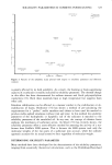
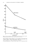
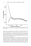
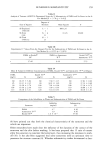
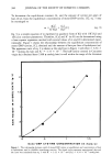
352 JOURNAL OF THE SOCIETY OF COSMETIC CHEMISTS 800- 800 400 go a'o INTERLAYER SPACING (•,) Figure 2. Comparison of x-ray diffraction spectra of washed and ether-extracted skin. /•, normal stratum corneum extracted for 30 minutes with ether ¸, washed normal stratum corneum O, region of complete spectral overlap. assigned with certainty. Tentatively, we interpret it as due to cavities formed in the structure by the extraction but are aware that other plausible explanations may exist. With this interpretation of the prominent feature of normal stratum corneum, the results are readily interpreted. First, washing of the separated stratum corneum removed epidermal lipids. From Figure 2, it can be seen that washing does not have as dramatic an effect on the stratum corneum as does extraction however, the effect on the 50- 80 • lipid region is virtually the same. The results gave essential information on the site of G2 in the stratum corneum. From the samples treated with G2 (Figure 3), it is evident that G2 interacts with and changes the structure of epidermal lipids. This is a relatively slow process continuing for several hours. Figure 3 reveals the trend of the change after one hour and also that the changes continue for a long time after that period. It is interesting to notice that the G2 dominated the spacings of the lipids. From earlier work on G2 (10), it was found that when G2 was added to a model skin-surface lipid mixture contained in a
Purchased for the exclusive use of nofirst nolast (unknown)
From: SCC Media Library & Resource Center (library.scconline.org)