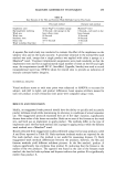
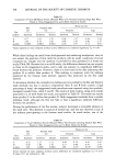
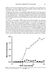
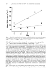
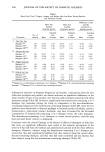
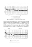
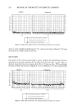
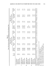
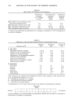
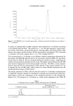
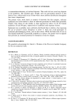
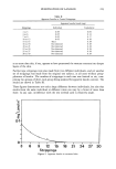
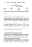
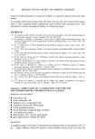
PENETRATION OF LANOLIN 221 The extent of any interference by steroids present in the natural intercellular lipids has been quantified and allowed for. GENERAL METHODOLOGY MATERIALS AND EQUIPMENT Scotch Magic © adhesive tape Glass fiber filter papers (Whatman GF/C, 11.0 cm) 55-mm glass funnels Glass rod, 120- x 5-mm flattened and flame-polished at one end 50-ml graduated stoppered glass cylinders Template, 20- x 10-mm rectangle cut in acetate sheet PROCEDURE The test subject was a 68-year-old male. A rectangular area 2 x 1 cm was marked out on the flexor surface of the inner forearm using a graphite pencil and the template. 4.2 mg of lanolin, weighed on a glass coverslip, were applied to this area and gently rubbed until absorbed, using the flame-polished glass rod. Ten minutes after application, tape stripping was commenced. Each stripping consisted of a 4-cm length of tape covering the test area, applied by thumb pressure for ten seconds and then removed by forceps. Successive strippings were collected in groups of three, each group being put into a 50-ml stoppered glass cylinder. To each cylinder 13.5 ml of chloroform were added, and after ten minutes the cylinder was shaken, causing the tape partially to disintegrate. 11.5 ml of acetic anhydride were then added, and the cylinder was reshaken, completing the disintegration of the tape and dissolution of any lanolin present. 0.5 ml of concen- trated sulphuric acid was added, another shake given, and the mixture immediately filtered through a glass fiber filter paper and filled into an optical cell. Simultaneously a blank was prepared from 12 cm of virgin tape, treated in a manner similar to the actual test. As the green color in the test mixture developed, the absorbance at 458 nm and 25øC was continuously monitored on the spectrophotometer until a maximum had been reached and just passed. The maximum reading was taken as definitive. The coverslip and glass rod used for lanolin application were washed with chloroform, and the residual lanolin thus recovered was determined by the same method as used for the tape strippings. The mass balance is shown in Table I, and essential results plotted as a graph are shown in Figure 1. The cumulative recovery oflanolin from all sources was 4.33 mg out of 4.20 mg applied (103.1%). Even though this recovery was excellent, since it was slightly in excess of the weight applied, it was necessary to ascertain to what extent natural steroids in the stratum corneum of the test subject had contributed to the measured amounts of lanolin. Accordingly, the whole experiment was repeated on the same subject, but this time without applying any lanolin. Faint positive reactions were found up to and including the fifth group of strippings, the results being shown in Table II and Figure 2. The

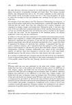
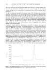
222 JOURNAL OF THE SOCIETY OF COSMETIC CHEMISTS Table I Recovery of Lanolin From Tape Strippings Lanolin found (mg) Strippings Individual Cumulative Cleanings 1.03 1.03 1 to 3 2.47 3.50 4 to 6 0.43 3.93 7 to 9 0.15 4.08 10 to 12 0.07 4.15 13 to 15 0.06 4.21 16 to 18 0.05 4.26 19 to 21 0.02 4.28 22 to 24 0.01 4.29 25 to 27 0.01 4.30 28 to 30 0.03 4.33 cumulative recovery of "lanolin" was 0.18 mg (note: this is not a constant figure see below), and after deducting this "blank" from the results of the original test with lanolin, the net recovery then becomes 4.15 mg out of 4.20 (98.8%). The weight of lanolin unaccounted for is only 50 Ixg, about the same as the limit of determination, and 3 m o 3 6 9 12 15 18 21 24 27 30 Strippings Figure 1. Lanolin removed by tape strippings.
Purchased for the exclusive use of nofirst nolast (unknown)
From: SCC Media Library & Resource Center (library.scconline.org)