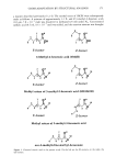
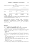
352 JOURNAL OF COSMETIC SCIENCE ing. Recently the harmfulness of ultraviolet (UV) radiation is increasing due to destruc- tion of the ozone layer. Excessive exposure to UV radiation causes postinflammatory pigmentation (4-5). Pigmentary disorders are caused by various factors, including in- flammation, the imbalance of hormones, and genetic disorder (6). Melanins play a critical role in the absorption of free radicals and melanogenesis in the skin in a kind of process that produces photoprotective agents against the damaging effect of UV. Many cosmetic and pharmaceutical companies have tried to find an inhibitor of melanogenesis. The regulation of cellular pigmentation can be controlled at many different stages of melanogenesis. Especially, tyrosinase inhibitors and antioxidants can be used for inhi- bition of cellular pigmentation since the melanin-producing process involves enzymatic and nonenzymatic oxidation reactions. Plant extracts having such biological activities may be a good choice for cosmetic purposes. We previously screened the inhibitory effects of 150 medicinal plants on elastase activ- ity, and examined their anti-inflammatory effects. The Areca catechu methanolic extract showed a high inhibitory effect on elastase and an anti-inflammatory effect, compared to reference compounds (7), and we selected the Areca catechu extract as a candidate for new anti-aging and anti-inflammatory agents. Areca catechu L. is widely cultivated, especially in southern Asia, and its seed is used as a chewing material anthelmintic and also as kompo-traditional medicine. Preparations containing Areca are also used as digestive medicines since Areca promotes the secretion of saliva (8-9). Areca catechu L. contains a number of chemical components, such as alkaloids, tannins, flavonoids, and fatty acids (10,11). To clarity the anti-aging mecha- nism of Areca catechu L. extract against aging, we have studied the anti-inflammatory effect in vitro and in vivo. Furthermore, the skin-whitening efficacy of the Areca catechu extract was also examined by inhibition of mushroom tyrosinase and inhibition of melanogenesis on B-16 melanoma cells. The anti-aging effect of Areca catechu L. extract was evaluated by measuring anti-oxidative activity, the free-radical scavenging effect, and inhibition of hyaluronidase in vitro, and anti-inflammatory effect/inhibition of de- layed hypersensitivity in vivo. The safety of CC-516 was evaluated by cytotoxicity on human fibroblasts and skin irritation testing. MATERIAL AND METHODS ANTIOXIDATIVE ACTIVITY A lipid peroxidation system was induced by Fenton's reagent. Each test sample (0.1 ml) and ethyl linoleate (10 pl) were added to an incubation medium (4.89 ml) containing 2% sodium dodecyl sulfate, 1 pM ferrous chloride, and 0.5 pM hydrogen peroxide. The known synthetic antioxidant, butylated hydroxytoluene (BHT) was used as a reference compound. The incubation medium was kept at 55 øC for 16 hr. Each reaction mixture (0.2 ml) was transferred into a test tube, followed by addition of 4% BHT (50 pl) to prevent further oxidation. Antioxidative activity of the sample was measured using a thiobarbituric acid (TBA) assay according to the method of Ohkawa eta/. (12). FREE-RADICAL SCAVENGING ACTIVITY Scavenging effect against free-radical generation was measured following the procedure

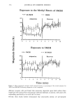
ARECA CATECHU L. EXTRACT 353 of Fugita et al. (13). The sample solution (2 ml) was added to 2 ml of 60 pM 1.1- diphenyl-2-picryl hydrazine (DPPH) ethanolic solution and kept at room temperature for 30 min. The absorbance was measured at 520 nm. ANTI-INFLAMMATORY INHIBITION OF HYALURONIDASE Hyaluronidase activity was determined spectrophotometically by measuring the amount of N-acetylglucosamine formed from sodium hyaluronate (14). Fifty microliters of bo- vine hyaluronidase (7,900 units/ml) dissolved in 0.1 M acetate buffer (pH 3.5) was mixed with 100 pl of a designated concentration of CC-516 dissolved in 5% DMSO, and then incubated in a water bath at 37øC for 20 min. The control group was treated with 100 pl of 5% DMSO instead of the CC-516. One hundred microliters of 12.5 mM calcium chloride was added to the reaction mixture, and then the mixture was incubated in a water bath at 37øC for 20 min. This Ca 2+ activated hyaluronidase was treated with 250 pl of sodium hyaluronate (1.2 mg/ml) dissolved in 0.1 M acetate buffer (pH 3.5), and then incubated in a water bath at 37øC for 40 min. One hundred microliters of 0.4 N sodium hydroxide and 100 pl of 0.4 M potassium borate were added to the reaction mixture, and then incubated in a boiling water bath for 3 min. After cooling to room temperature, 3 ml of dimethylaminobenzaldehyde solution (4 g of p-dimethylamino- benzaldehyde dissolved in 350 ml of 100% acetic acid and 50 ml of 10 N hydrochloric acid) was added to the reaction mixture, and then incubated in a water bath at 37øC for 20 min. Optical density at 585 nm of the reaction mixture was measured by using a spectrophotometer. The percentage of inhibition was calculated as: Inhibition (%) = [(ODc-ODs)/OD c] x 100 where ODe is the OD at 585 nm of the control, and ODs is the OD at 585 nm of the sample. ANTI-INFLAMMATORY ACTIVITY For measuring the topical anti-inflammatory activity, the mouse ear edema assay was employed. According to the modified method of Tonnel et al. (15), preparations of the CC-516 were topically applied to the right ears of mice (18-22 g) three times at 3-hr intervals. Thirty minutes after the final treatment of the test compounds, 2.5% croton oil or 2% arachidonic acid dissolved in acetone (25 lal/ear) was applied topically to the ears of the mice, and the ear thickness was measured 5 hr after croton oil treatment or 1 hr after arachidonic acid treatment. Percent inhibition of ear edema was calculated by comparison with the control group having the vehicle and anti-inflammatory only. Inhibitory activity against delayed hypersensitivity was measured according to the method of Tarayre et al. (16). Briefly, 3% picryl chloride (acetone) was applied to the abdomen of mice (18-22 g). One week later, 3% picryl chloride was applied to the ears of the mice, and ear thickness was measured 24 hr after the picryl chloride solution treatment. Preparations of the test compounds were applied to the ears of the mice daily for 7 days starting from day 0. The differences between the ear thickness of the extract- treated group and the control group treated with picryl chloride and vehicle only were regarded as an indication of inhibitory activity.
Purchased for the exclusive use of nofirst nolast (unknown)
From: SCC Media Library & Resource Center (library.scconline.org)