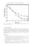
PLANT SMALL RNA TECHNOLOGY 119 CELL CULTURE Fibroblasts were extracted from different normal human skin samples obtained from do- nors and were grown in Dulbecco’s modifi ed eagle medium (DMEM) 1 g/L glucose (Lonza, Verviers, Belgium) supplemented with 10% of FBS (Lonza), 2 mM of L-Glutamine (Lonza), and 100 μg/ml of Primocin (InvivoGen, San Diego, CA). Cell cultures were maintained at 37°C in a 5% CO2-humidifi ed atmosphere. In the model of replicative senescence, fi broblasts were cultured over a 3-mo period. At each passage, when cells reached confl uence, a part of these cells were frozen and the other part was maintained in culture. After different passages (P-X), the cells were thawed and treated, or not (control condition), overnight or for a period of 24 or 48 h with 1% PSR baobab extract or with 1% placebo (baobab extract without small RNAs) (twice a day). HUMAN SKIN SAMPLES Ex vivo skin samples were obtained from donors after undergoing abdominal plastic sur- gery. On skin biopsies of 6 mm in diameter, a topical application of 20 μl of either 1X phosphate-buffered saline (PBS) (control) or 1% PSR baobab extract or 1% placebo di- luted in 1X PBS (baobab extract without small RNAs) was applied twice a day for 48 h. Then, tissues were fi xed in formalin and embedded in molten paraffi n wax. qPCR After treatment, the culture medium was removed and the cells were rinsed with cold PBS. The mirVana miRNA Isolation Kit (Ambion, Naugatuck, CT) was used to extract total RNAs. Then, these RNAs were reverse-transcribed with the High-Capacity cDNA Reverse Transcription Kit with RNase inhibitors (Applied Biosystems, Foster City, CA). The reverse transcription was performed on 0.25 μg of total RNA using a MiniCycler thermocycler (MJ Research, Waltham, MA). Finally, real-time PCR was performed on a StepOnePlus thermocycler (Applied Biosystems). Collagen I, miRNA-19b or Drosha TaqMan Gene Expression Assay (Applied Biosystems) and TaqMan Gene Expression Master Mix (Applied Biosystems) were involved. RNU44 TaqMan Gene Expression As- say was used as an endogenous control to miRNA expression studies and 18S TaqMan Gene Expression Assay to Collagen I and Drosha expression study. The comparative Ct method was used for relative quantifi cation of target expressions (17), and the StepOne* Software (Applied Biosystems) was used for data treatment. SENESCENCE-ASSOCIATED (SA) β-GALACTOSIDASE STAINING Fibroblasts were washed in PBS, fi xed for 3 min in 2% formaldehyde and 0.2% glutaral- dehyde solution, washed, and incubated overnight at 37°C with SA β-galactosidase stain solution at 1 mg/ml [MgCl2 at 2 mM, K3Fe(CN)6 at 5 mM, K4Fe(CN)6 at 5 mM, NaCl at 150 mM, citric acid at 40 mM, and X-gal (Qiagen, Hilden, Germany)]. The slides were rinsed and mounted in Aquatex (Merck, Kenilworth, NJ). Detection was managed and examined using a Nikon Eclipse E600 microscope with a ×20 objective. Photos were taken with a QImaging* EXi blue camera and processed by using the Q-Capture Pro 7 (QImaging, Survey, BC, Canada) acquisition software.

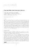
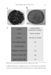
JOURNAL OF COSMETIC SCIENCE 120 For image quantifi cation, three images per condition were analyzed. Photoshop Elements 11 software (Adobe, San Jose, CA) allows users to convert each picture into an inverted image, using an intensity threshold to eliminate the background noise. These inverted images were then analyzed with Volocity image analysis software (Improvision), which generated the sum of red pixel intensities. The sum obtained was normalized by taking into account the total number of cells. IMMUNOFLUORESCENCE STAINING OF COLLAGEN I Sections were deparaffi nized and rehydrated with several successive xylene, alcohol, and water baths. Then, an unmasking protocol was performed that included both microwave exposure at 600 W in citrate buffer 0.01 M, pH 6 (Sigma, Saint Louis, MO) until boiling and 0.05% trypsin (Zymed, Invitrogen, Frederick, MD) digestion for 15 min at 37°C. The saturation of unspecifi c sites was performed with a solution of 5% bovine serum al- bumin (BSA) (Sigma) for 30 min. Depending on the experiment, the primary antibody used corresponded to polyclonal anti-collagen I (Rockland at 1:100 dilution in 1X PBS). After rinsing the slides with PBS, the secondary antibody was applied in the dark and under agitation at room temperature in a damp room. Finally, the sections were mounted in Fluoromount-G (Electron Microscopy Sciences, Hatfi eld, PA). Detection was managed and examined using a Zeiss Axiovert 200M microscope with a ×20 objective. Photos were taken with a QImaging EXi blue camera coupled to Volocity acquisition software (Improvision). STATISTICAL ANALYSIS Statistical analyses were performed using JMP* 10 software (SAS, Carry, NC) and Stu- dent’s t test for independent samples with one-tailed direction of rejection. p 0.05 was considered as signifi cant, p 0.01 as very signifi cant and p 0.005 as highly signifi cant. RESULTS/DISCUSSION COMPOSITIONAL ANALYSIS OF BAOBAB SEEDCAKE The baobab seedcake was ground before raw material analysis to obtain a powder around 2,000 μm in diameter (Figure 2B). Then, the chemical composition of the baobab seed- cake was studied. Phytochemical analysis of the baobab seedcake revealed that the seed- cake was very rich in different classes of chemical molecules (Figure 2C). This confi rms its potential for use as a raw material on which to perform an aqueous extraction and obtain a biofunctional ingredient for use in the cosmetic fi eld. COMPOSITIONAL ANALYSIS OF BAOBAB SEEDCAKE EXTRACTS The composition of the baobab seedcake extracts was studied. The small RNA content in both the target extract and the placebo extract was determined by bioanalyzer analysis (Figure 3). A high amount of small RNAs was found in the extract obtained by the spe- cifi c patented process intended to enrich the extract in small RNAs, around 60 mg/l be- fore dilution. By contrast, the extract obtained without PSR technology process did not contain small RNAs at all.
Purchased for the exclusive use of nofirst nolast (unknown)
From: SCC Media Library & Resource Center (library.scconline.org)