DEAD SEA MUD & CUTANEOUS BLOOD FLOW 261 mixed with distilled water were called “As is” mud and the one mixed with Dead Sea water was called “Salted” mud. Then, both were autoclaved for 15 min at 121 C. Samples were sent to the Royal Scientifi c Society Instrumental Analysis Laboratories (Amman, Jordan) for analysis of their salts and element contents. Laboratory tests and analyses performed included moisture content loss on ignition (BS EN 196-2) total Al, K, Ca, Cr, Co, Ni, Cu, Zn, As, Cd, Pb, Li, Sr, Na, Mg, Mn, Fe, and SiO2 (SOP No. 3/01/04-005, atomic absorption spectroscopy) total Hg and As (SOP No. 3/01/04-006, atomic absorption spectroscopy) total Br (ASTM D-3869-79, gravimetric analysis) and Cl water soluble (BS EN 196-2). SUBJECTS Fifteen healthy, normotensive, nonsmoking female volunteers aged 18–45 years partici- pated in the study after giving their informed consent. Subjects with any lesions on their forearms or receiving any local or systemic treatments were excluded from the study. The study protocol was offi cially approved by the Hashemite University Institutional Review Board. Subjects were randomly assigned to either “Salted” mud group (n = 5), “As is” mud group (n = 5), or “Over-the-Shelf” mud group (n = 5). Each subject served as her own control. INSTRUMENT Skin blood fl ow and temperature were monitored pre-, during, and postmud application using moorVMS-LDF dual channel—laser Doppler and temperature monitoring (Perimed® PF4001, wavelength 82 nm, Perimed, Moor, UK) and recorded by the system software with a 3-s time constant downstream from a broadband fi lter (12 MHz). Laser Doppler is a gold standard for dynamic microvascular blood fl ow assessment. Laser Doppler fl owmetry (LDF) provides continuous noninvasive measurements of microvascu- lar perfusion in terms of fl ux, which represents the movement of erythrocytes through the microvasculature (10). It was used to assess the effect of local treatment in improving skin microcirculation, thus improving the healing of skin ulcers (11) and to assess the effect of a topical product on microcirculation (12). In addition, LDF data correlate well with vi- sual scores of patch test reaction and is used in dermatology to predict irritancy and to assess topical products such as anti-infl ammatory, vasoactive drugs, and detergent barrier creams (10). A dual channel laser Doppler monitor (VMS-LDF2) with two (2) laser probes for simul- taneous recording of skin microcirculation and skin temperature was used. VMS-LDF2 was computer-controlled with software (MoorVMS-PC V2.2). The sampling depth of the probe is around 1 mm. Low-power laser light is transmitted via the optic probe to the tissue, and the light is scattered by the tissue and moving blood cells and their frequency is Doppler-broadened. Some of the scattered light is collected by the optic probe and transmitted to a photodetector. The data are electronically processed by the system software to produce the laser Doppler (blood fl ow) fl ux signal. The probes were calibrated just before the start of the study according to the manufacturer’s guidelines. Perfusion values were measured for each tested time points at a sample rate of 33 recordings per second

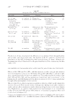
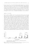
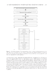
JOURNAL OF COSMETIC SCIENCE 262 and were analyzed by averaging over 1-min intervals for protocol 1 and averaging over 10 min before and 30 min during intervals for protocol 2 using the manufacturer software. STUDY DESIGN The participants were randomly assigned into three groups of fi ve subjects each. Each group (n = 5) was used to test one of the aforementioned mud types. According to the published guidelines for measurement of cutaneous blood fl ow (13), the subjects were asked not to drink caffeinated beverages, not to eat a high-calorie meal at least 2 h before the experiment, and not to exercise at least 24 h before the experiment. The subjects were asked to relax for 20 min in a supine position while their forearms are relaxed and in an extension position in a controlled temperature and a controlled relative humidity room. Lighting conditions in the room were kept constant by closing window blinds and turn- ing off ceiling lights, and there was typically suffi cient light to be able to read. Five cir- cular areas were marked on the ventral aspect of each forearm (Figure 1). One forearm was Figure 1. Five circular areas were marked on the ventral aspect of each forearm. Areas 1–4 were used for protocol 1 and area 5 for protocol 2. For protocol 1 laser Doppler readings (fl ux and temperature) were taken for areas 1–4 at baseline before mud application and then directly and 15 min after mud removal.
Purchased for the exclusive use of nofirst nolast (unknown)
From: SCC Media Library & Resource Center (library.scconline.org)