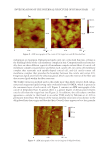
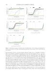
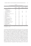
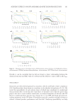
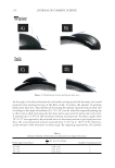
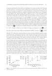
INVESTIGATION OF THE INTERNAL STRUCTURE OF HUMAN HAIR 119 Young’s modulus) of various lamellar constituents of the cuticle (27,28). Other studies focused on elucidating changes in the nanomechanical properties as a result of bleach- ing treatments or by changing the relative humidity. Not surprising, both bleaching and exposure to increasing levels of relative humidity reduce the modulus of the cuticle (29,30). As technology in the fi eld of AFM progresses, so does the analysis of hair by a variety of novel techniques. For example, surface potential imaging experiments—also known as Kelvin probe microscopy—were carried out to determine the electrostatic properties of the hair surface (31–34). Key fi ndings from these studies shed light on the surface poten- tial properties of the cuticle, in particular demonstrating the increased polar nature of the cuticle edge and also the infl uence of lipids on the wettability properties of the fi ber (31,32). In addition, a nanoscale understanding of the electrostatic (static charging) prop- erties of hair was also garnered with Kelvin probe microscopy (33,34). Other important contributions by AFM technologies to understanding phenomena in hair include the use of nanoscale infrared spectroscopy and imaging for the high-resolution localization of structural lipids in hair (35). New insights have also been gained by attach- ing individual hair fi bers to AFM probes and measuring the interaction between the probe fi ber and another hair fi ber (36,37). Furthermore, one of the caveats of any micros- copy technique with human hair is the diffi culty of monitoring the same region before and after treatment, which is circumvented with a practical mounting solution offered by Breakspear and Smith (38). Almost all of the AFM studies investigating human hair have focused on probing the outer surface of the cuticle. From a different perspective—using torsional resonance mode AFM—researchers at Ohio State University analyzed cross sections and transverse sec- tions of human hair (39,40). These studies shed light on some of the internal structures of hair, and the researchers were able to differentiate between the different sublamina of the cuticle and several features of the hair cortex. In this article, we demonstrate how a cross-sectioning technique in combination with contact-mode AFM allows us to visualize the various ultrafi ne structural components of the cuticle and cortex and investigate the infl uence of bleaching and delipidation on these structures. MATERIALS AND METHODS Studies were conducted on Asian hair purchased from International Hair Importers & Products Inc. (Glendale, NY). Before analysis, all hair was washed with a 3% (w/w) sodium laureth sulfate:cocamidopropyl betaine (12:2) mixture. BLEACHING OF HAIR Bleaching was carried out by mixing 120 g of Clairol Professional BW2 powder lightener (The Wella Corporation, Woodland Hills, CA) with 147 mL of Salon Care Professional 20 Volume Clear developer (Arcadia Beauty Labs LLC, Reno, NV). The resulting mixture was applied to damp hair for two 30-min intervals for a total bleaching time of 1 h. Bleached hair was shampooed twice with 3% (w/w) sodium laureth sulfate:cocamidopropyl betaine (12:2) before experiments.

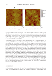
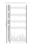
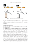
JOURNAL OF COSMETIC SCIENCE 120 DELIPIDATION OF HAIR Using a Soxhlet extraction apparatus, free internal and surface lipids were removed from hair. This method is based on an established procedure in which hair is extracted with a series of solvents of increasing polarity (41). The apparatus consisted of a round-bottom fl ask to which a Soxhlet extraction tube was mounted. Inside the Soxhlet extraction tube, a bundle of hair was placed in a cellulose thimble. A condenser was mounted on top of the Soxhlet extraction tube. The effect of solvent extraction on hair was investigated fi rst by treatment with t-butanol and n-hexane for 4 h each, then with a mixture of chloroform/ methanol (70:30, v/v) for 6 h. In each procedure, 3 g of hair was treated with 250 mL of solvent in the Soxhlet extractor. PREPARATION OF HAIR CROSS SECTIONS Hair fi bers were embedded in an epoxy resin block. The hair-epoxy block was cut into approximately 2-mm-thick sections. A Buehler MetaServ 250 Grinder-Polisher (Lake Bluff, IL) was used to prepare a smooth surface of the hair cross sections, suitable for AFM analysis. In this procedure, the hair-epoxy sections were sequentially grinded with alumi- num oxide sandpaper with grit levels of 300, 600, and 1,200 with a head force set to 0.5 lb (|2.22411 N). Both the head and plate rotate at a speed of 150 rpm in opposite directions. The lower head force and spin rate ensure mild abrasion and no tearing of the hair specimen. After sandpaper treatment, polish was applied with a liquid suspension of Al2O3 (0.05 μm). Suspended Al2O3 particles can roll and gently slide across the cloth and specimen. Finally, the hair-epoxy section was sonicated in deionized water for 1 min to eliminate the residues generated from grinding and polishing. This polishing technique was chosen as an alternative to conventional microtome preparation, which can introduce a variety of artifacts, including scores and tearing, chatters (thick and thin zones parallel to the knife edge), and compression artifacts. If performed incorrectly (e.g., with a large head force or high spinning speed), grinding and polishing can also introduce artifacts to the sample resulting in scratches, tearing, and uneven surfaces. Polishing is a preferential technique for preparing various biological specimens, such as bone or tooth, for micro- scopic investigations. Because a rough surface prevents the AFM probe from being able to raster effectively over the sample, the polishing technique creates a smooth surface and makes the sample more amenable for AFM analysis. AF M MEASUREMENTS Co ntact-mode AFM studies were carried out using a Multimode Nanoscope III (Bruker Corporation, Billerica, MA) using a 128-μm piezoelectric scanner. In this technique, a small tip attached to a cantilever is rastered in the X and Y directions on the surface of a sample, and the height (Z direction) is monitored. The operation of the instrument is based on the principal that a diode laser beam is focused on the top of a cantilever and refl ected onto a position-sensitive photodetector. The probe (cantilever and tip) is shaped like a stylus, and as it traverses across the surface, the topographic features are mapped based on forces encountered between the probe and the sample. An Si3N4 probe (SNL-10) with a triangular cantilever (force constant of 0.06 N/m) from Bruker Corporation was used for all experiments. We operated the instrument in constant force mode, in which the
Purchased for the exclusive use of nofirst nolast (unknown)
From: SCC Media Library & Resource Center (library.scconline.org)