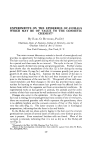
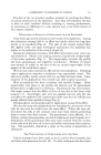
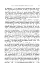
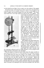
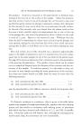
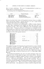
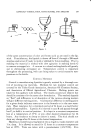
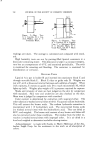
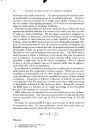
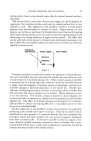
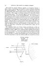
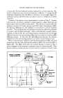
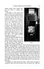
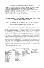
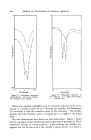
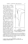
EXPERIMENTS ON EPIDERMIS OF ANIMALS 231 The skin of the rat provides excellent material for studying the effects of various substances on the epidermis. Just what will stimulate the skin to have an ideal condition without irritating it, causing parakeratosis, an acanthosis, or affecting it in some adverse way is the chief interest of the cosmetic chemist. HISTOLOGICAL EFFECTS OF SUBSTANCES ON THE EPIDERMIS A few years ago several substances were tested on the epidermis. Among the substances causing little or no effect was lanolin which has long been known to be beneficial (Fig. 4). The skin of the treated animal often felt slightly softer and upon histological examination the epidermis was similar to the epidermis of the control animal (4). Among the subs.tances causing a mild effect were stearic acid, castor oil, and mineral oil. Much to my surprise, mineral oil produced a hypertrophy of the entire epidermis (Fig. 5). This hypertrophy involved the prickle cell layer, granulosum, and imperfect cornification. Perlman (5) found that mineral oil added to the diet of the rat caused hypertrophy of the gingiva of the mandibular region. Olive oil and xylene extensively affected the entire epidermis. Following xylene applications imperfect cornification was particularly noted. The cells were swollen, loosely united with air, and fluid between them. Upon cessation of the applications the corneum was shed in great quantities. Olive oil produced the most consistent and marked changes. Great hypertrophy took place in the prickle cell stratum and the granulosum became seven or eight layers in thickness. Parakeratosis was very marked. One might suspect that the effect of olive oil was due to free fatty acids present in it. Accordingly, oleic acid was applied and not only did this acid affect the epidermis but it penetrated down into the hair follicles and greatly affected their epithelial lining (Fig. 6). Ethylene glycol and propylene glycol applications caused little effect. Oils did not cause the parakeratosis by retarding the desiccation of the cells for the wool fat would have the same effect. Where oils were ad- ministered the cells of the corneum must have been altered, permitting them to retain their fluid content. The granulosum is also affected. It is thicker, contains more granules, and mitotic figures are more frequent in it. Stimulation of the granulosum cells must have resulted from the contact of the cells with the olive oil or oleic acid which necessitates their penetra- tion. This possibility stimulated a study on the extent of penetration of substances. THE PENETRATION OF SUBSTANCES INTO THE SKIN Various attempts have been made to see if, and by what channels, fats and fatty acids penetrate the skin (6). In other techniques fats have been

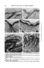
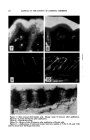
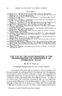
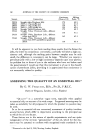
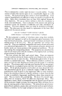
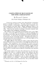
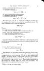
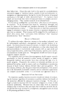
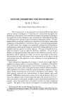
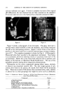
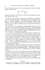
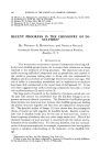
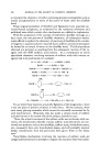
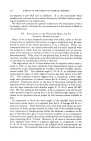
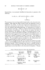
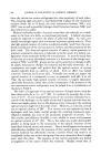
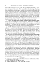
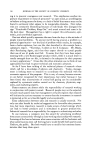
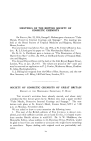
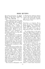
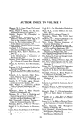
232 JOURNAL OF THE SOCIETY OF COSMETIC CHEMISTS ?• :- ...... • ..•:• ......... •. ½:-•:•.:• •.• :•.•:..• .... •-,,•, ..•., •.• •:• • . . • .. ....... •:• • . . • •.•::.: •. .,•:•,, :•:.., .... '•,• .... • .•-•. •*• . ,•-.-½-:.-..•-¾ ":=: ::8.• •': ß :•½:• .•.• 4•g•. -'. .•'•*":'.,•'• ':'*" ":' •"•,•'•'•.•% ß •Z'.•" ' . •.... .., ½•4 "" ...... - % -.4' ,, . .....¾½.'-?" ,• • . ':::. Figure 7.--Skin treated with linoleic acid. Biopsy taken 10 minutes after application. Figure 8.--Lanolin 10 minutes after application. Figure 9.•Normal skin of rat. Figure 10.--Biopsy of skin 20 minutes after application of linoleic acid. Figure 11.--Applications of iodinated linoleic acid were applied at 9:30, 11:30, and 1:30• and four hours later the biopsy was taken.
Purchased for the exclusive use of nofirst nolast (unknown)
From: SCC Media Library & Resource Center (library.scconline.org)