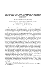
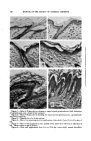
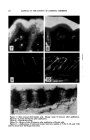
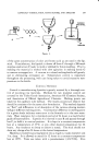
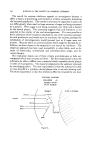
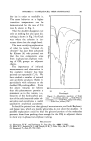
EXPERIMENTS ON EPIDERMIS OF ANIMALS 233 applied to the skin and the skin stained after sectioning to obtain the loca- tion of the fat (7). In either instance, the dyes spread to adjacent tissues and no one has ever been able to demonstrate fatty acid penetration or destination too clearly (8). Theoretically some lipoid substances such as cholesterol, lecithin, and the fatty acids should be able to penetrate since they are miscible to some extent in both fats and water. The penetration of fats and fatty acids was therefore studied by fluorescence. They pos- sessed or enhanced fluorescence due to substances dissolved in them (9). Applications of the substances were made, biopsies were taken, fixed in formalin, and sections were cut on the freezing microtome. Linoleic acid penetrates the epithelium rapidly (Fig. 7). Twenty minutes after the application sections show that linoleic acid is present in the blood vessels (Fig. 10). Cross sections of various vascular channels show that linoleic acid adheres as a thin film to their lining. Oleic acid is also absorbed readily. Droplets of varying sizes can be seen in the epidermal cell layers ten minutes after the application. Only minute amounts are ever found in the blood vessels at any time, indicating that passage into the vessels is slow, not extensive, or there is little retention in the vessels. Lanolin (Fig. 8) and ricinoleic acid were retained mainly in the outer strata of the epidermis. If they penetrate their absorption must be very slow or in small amounts since they cannot be detected by fluorescence. The path of penetration of substances through the skin has been thought to be via the hair follicles. Since the linoleic acid is found in the horizontal plexus of vessels under the epithelium before it is found in vessels around the sebaceous glands, a great amount must pass directly through the epidermis. The linoleic acid must affect the epithelial cells and decrease their pro- tective properties since after several applications the acid seems to pene- trate faster and in greater quantities. The penetrating fatty acids prob- ably induce growth and repair of the epithelium as does any other injury. The fact that lanolin was retained by the superficial strata of the epi- dermis and did not penetrate more deeply is surprising. However, lanolin did not penetrate in the experiments of others (6, 10). Radioactive sodium has also been absorbed from fatty bases (11). While the present experiments on penetration were quite convincing and it appeared that linoleic acid even entered the blood vessels, this observa- tion needed confirmation. Radioactive linoleic acid was sought with the intention of following its course through the epithelium. However, such acid could not be obtained and linoleic acid was iodinated converting it into mono-iodo-stearic acid. Applications of the iodinated material were made, biopsies were taken, and slides prepared (12). Great penetration took place into the hair folli-

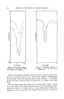
234 JOURNAL OF THE SOCIETY OF COSMETIC CHEMISTS cles and sebaceous glands and some penetration through the epidermis directly (Fig. 11). The amount in the dermis seemed to depend upon the quantity applied, either by means of concentration or number of applica- tions. The dermis acted as a barrier and restricted the depth of penetra- tion. Several days after the last application, the material in the dermis had diffused and much of the epidermis had shed, the shedding cells carrying with them the radioactive material. There was no evidence that the iodi- nated material had passed in any quantity into the blood vessels. Iodination of the linoleic acid molecule probably prevented such passage. The effect of the radioactive material on the N. B. T. plates was so diffuse that one could not determine whether the material passed through or be- tween the cells of the epidermis. Likewise the exact depth of penetration could not be determined. This was disappointing and a better method needs to be devised. Throughout these penetration studies, it was noted that many of the fatty acids were much more effective at a certain time in the hair cycle. For instance, when the hair coat is resting, there is much penetration, and as the time of growth is approached, applications of the fatty acids are less irritating. This led to a detailed study of the skin and the cause or causes of these different effects. FLt•ID P^ss^GE T.Rot•G. T,E SKttq Since the application of fatty acids was more irritating at some intervals than others, the fluid condition of the epidermis was suspected as making these effects possible. One means of investigating the fluid aspect was to determine the fluid passage through the epidermis. For determining fluid loss, skin of different aged animals was stretched across diffusion chambers (13) containing 10 cc. normal saline. The cham- bers were inverted and left on a screen in an oven maintained at 35øC. and at a humidity of 28-32 per cent. The fluid loss through the skin of rats 22 days old averaged 1.302 mg./sq. cm. for the first hour. The loss gradually increased and by the 29th day of life the average loss per square centimeter was 2.922 mg. for the first hour. Fluid loss through the epidermis is therefore least when the epidermis is thinnest, there is no distinct granulosum and the comeurn is dry, hard, and brittle. There is less fluid in the skin as shown by a previous investigation (3) and more is evaporated than is supplied by the underlying tissues. This tends to dry out the corneum which aids in retarding and reducing the fluid loss. In the 30-day-old rat the epidermis is thicker, and a distinct granulosum is present. At this age the fluid content of the skin is greater. The ratio of the fluid supplied the epidermis by the underlying tissues in respect to evaporation is greater than in a 22-day-old rat and thus a moist comeurn exists.
Purchased for the exclusive use of nofirst nolast (unknown)
From: SCC Media Library & Resource Center (library.scconline.org)