1,4-DIOXANE ASSAY 213 (15) (16) (17) (18) (8) S.C. Rastogi, Headspace analysis of 1,4-dioxane in products containing polyethoxylated surfactants by GC-MS, Chromatographia, 29, 441-445 (1990). (9) S. Scalia, M. Guarneri, and E. Menegatti, Determination of 1,4-dioxane in cosmetic products by high-performance liquid chromatography, Analyst, 115, 929-931 (1990). (10) M.P. Italia and M. A. Nunes, Gas chromatographic determination of 1,4-dioxane at the parts-per- million level in consumer shampoo products, J. Soc. Cosmet. Chem., 42, 97-104 (1991). (11) C. Hoch-Ligeti, M. F. Argus, and J. C. Arcos, Induction of carcinomas in the nasal cavity of rats by dioxane, Br. J. Cancer, 24, 164-167 (1970). (12) National Cancer Institute, "Bioassay of 1,4-Dioxane for Possible Carcinogenicity," in Technical Report Series No. 80, NCI-CG-TR-80, U.S. Dept. of Health, Education and Welfare (Washington DC, 1978). (13) F. N. Marzulli, D. N. Anjo, and H. I. Maibach, In vivo skin penetration studies of 2,4-toluene- diamine, 2,4-diaminoanisole, 2-nitro-p-phenylenediamine, p-dioxane and N-nitrosodiethanolamine in cosmetics, Food Cosmet. Toxicol., 19, 743-747 (1981). (14) International Agency for Research on Cancer, "l,4-Dioxane," in Monographs on the Evaluation of Carcinogenic Risk of Chemicals to Humans, World Health Organization (Lyon, France, 1976), Vol. 11, pp. 247-256. H. W. Leung and D. J. Paustenbach, Cancer risk assessment for dioxane based upon a physiologi- cally-based pharmacokinetic approach, Toxicol. Lett., 51, 147-162 (1990). European Economic Community Council Directire 76/768/EEC, Appendix II (1976). C. F. Poole and S. A. Schuette, Contemporary Practice of Chromatography (Elsevier, Amsterdam, 1984), pp. 156-159. C. F. Poole and S. A. Schuette, Contemporary Practice of Chromatography (Elsevier, Amsterdam, 1984), pp. 607-608.

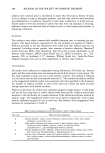
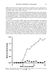
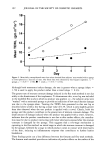
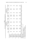
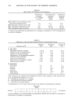
j. Soc. Cosmet. Chem., 43, 215-217 (July/August 1992) Iron content of human epidermis from sun-exposed and non-exposed body sites DONALD L. BISSETT and JAMES F. McBRIDE, The Procter & Gamble Company, Miami Valley Laboratories, Cindnnati, OH 45239-8707. Received March 3, 1992. Synopsis In previous work we observed that the iron content of human dermis is greater in sun-exposed than in non-exposed body sites. The present study indicates a similar difference in iron content for epidermis from these body sites. We speculate on the possible role of the iron in epidermal photodamage. INTRODUCTION Chronic exposure of hairless mice to UVB (1) or UVC (2) has been shown to result in an increase in non-heme iron content of the exposed skin. Analysis of human skin from sun-exposed vs non-exposed body sites also revealed a greater content of iron in the former (1). Based on histological and immunohistochemical analyses (1), the iron in UV-exposed mouse and human skins appeared to be present primarily as large dermal deposits. Iron can participate in generation of oxygen radicals (3-6), which are dam- aging to biological targets such as protein, lipid, and nucleic acid (6-11). The increased iron in UV-exposed skin would increase the likelihood of damage via this mechanism. The observation that topical iron chelators are photoprotective in mice (1) suggests that this mechanism is a significant factor in photodamage. Since topical chelators were protective against damage to both dermal and epidermal components (1), it is likely that substantial levels of iron are present in the epidermis, as they are in the dermis. In the present study, iron analysis of human epidermis was done to measure the basal level of this element in skin from non-exposed sites and, as was observed previously for the dermis, to determine if the content is greater in sun- exposed sites. MATERIALS AND METHODS HUMAN EPIDERMIS Human skin was obtained from remaining frozen samples of the same donor skins described previously (1). Donors were two males and six females, ages 53 to 80 years. 215
Purchased for the exclusive use of nofirst nolast (unknown)
From: SCC Media Library & Resource Center (library.scconline.org)