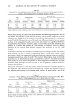
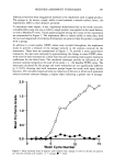
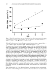
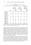
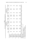
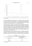
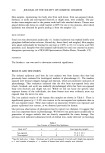
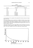
216 JOURNAL OF THE SOCIETY OF COSMETIC CHEMISTS Skin samples, representing two body sites from each donor, from sun-exposed (cheek, forehead, or neck) and non-exposed (buttock or thigh) areas, were available. The par- ticular tissue samples used in the present work had little attached dermis. Tissues were placed dermis side down on a slide warmer at 60øC for one to two minutes, after which epidermis was obtained by gently peeling it from the samples (12). IRON CONTENT Total iron was determined analytically (1). Isolated epidermis was washed briefly with phosphate-buffered saline solution, blotted dry, freeze dried, and weighed. Skin samples were ashed individually by heating for one hour at 210øC in 10:1 (v:v) nitric acid:70% perchloric acid. Samples were then assayed individually for total iron content by atomic absorption spectroscopy in a PE3030B Spectrometer (Perkin-Elmer, Norwalk, CT). STATISTICS The Student t test was used to determine statistical significance. RESULTS AND DISCUSSION The isolated epidermis used here for iron analysis was from human skin that had previously been evaluated for histological markers of photodamage (1). The markers assessed were dermal collagen damage and elastosis, which are prominent indicators of photodamage. That evaluation indicated that the skin from sun-exposed body sites (cheek, forehead, and neck) was markedly photodamaged while that from non-exposed body sites (buttock and thigh) was not. While we did not know the specific solar exposure history of the individuals, the three former sites were evidently more sun exposed than the latter two sites. The iron analysis results of the human skin samples are shown in Table I. There is significantly greater (p 0.05) iron content in the sun-exposed site epidermis than in the non-exposed tissue. These data indicate an association between sun exposure and greater epidermal iron content, as we observed previously for dermis. Our previous observations of photoprotection by topical iron chelators (1) suggest that iron is an important factor in photodamage. We believe iron participates catalytically in generation of oxygen radicals that are directly responsible for tissue damage. Our previous work indicated substantial levels of dermal iron, easily detected via histology Table I Iron Content of Human Skin Epidermis Body site* n n' Iron (ppm dry weight) Sun-exposed 7 7 53.0 -+ 34.3 Non-exposed 9 8 17.8 + 9.3 * Specific body sites: 1 cheek, 3 forehead, and 3 neck (sun-exposed) 3 buttock and 6 thigh (non-exposed) n = total number of samples n' = number of donors from which samples were obtained.

IRON CONTENT OF EPIDERMIS 217 or immunohistochemistry as localized deposits. That work did not reveal any deposits in the epidermis. The methods used, though, were not particularly sensitive to low levels of iron, which would occur if there were a more uniform distribution throughout that tissue compartment. The present work, while based on analysis of relatively few skin samples, indicates epidermal iron levels similar to those we observed previously for whole skin (primarily dermis). Our values are also similar to those reported by others (13,14) for human epidermis (26 and 98 ppm, respectively) of undefined solar exposure history. Epidermal iron is probably more uniformly distributed than in dermis, thus requiring the use of sensitive atomic absorption to detect it. This iron could participate in epidermal photodamaging events, such as skin cancer. While the basal level of iron in non-exposed epidermis is potentially available as a catalyst, the greater level in exposed skin provides an increased potential for formation of reactive oxygen. ACKNOWLEDGMENT We gratefully acknowledge Dr. David L. Windsor of the Procter & Gamble Company for his iron analytical work. REFERENCES (1) (2) (3) (4) (5) (6) (7) (8) (9) (lO) (11) (12) (13) (14) D. L. Bissett, R. Chatterjee, and D. P. Hannon, Chronic ultraviolet radiation-induced increase in skin iron and the photoprotective effect of topically applied iron chelators, Photochem. Photobid., 54, 215-223 (1991). S. S. Ranade, P. Murugaiyan, B. S. Manerikar, and S. D. Joshi, Alteration of macromolecular events and elemental levels in the skin of UVC exposed hairless mice, Physiol. Chem. Phys. Med. NMR, 18, 197-205 (1986). G. R. Buettner, Activation of oxygen by metal complexes and its relevance to autoxidative processes in living systems, Bioelectrochem. Bioenerg., 18, 29-36 (1987). • H. B. Dunford, Free radicals in iron-containing systems, Free Rad. Biol. Med., 3, 405-421 (1987). A. Puppo and B. Halliwell, Formation of hydroxyl radicals from hydrogen peroxide in the presence of iron, Biochem. J., 249, 185-190 (1988). G. Minotti and S. D. Aust, The role of iron in initiation of lipid peroxidation, Chem. Phys, Lipids, 44, 191-208 (1987). J. M. Braughler, L. A. Duncan, and R. L. Chase, The involvement of iron in lipid peroxidation, importance of ferric to ferrous ratios in initiation, J. Biol. Chem., 261, 10282-10289 (1986). K. J. A. Davies, Protein damage and degradation by oxygen radicals. I. General aspects, J. Biol. Chem., 262, 9895-9901 (1987). S. Inoue and S. Kawanashi, Hydroxyl radical production and human DNA damage induced by ferric nitrilotriacetate and hydrogen peroxide, Cancer Res., 47, 6522-6527 (1987). M. J. Peak and J. G. Peak, Hydroxyl radical quenching agents protect against DNA breakage caused by both 365-nm UVA and by gamma radiation, Photochem, Photobid., 51, 649-652 (1990). H. C. Schroder, R. Messer, M. Bachmann, A. Bernd, and W. E. G. Muller, Superoxide radical- induced loss of nuclear restriction of immature mRNA: A possible cause for aging, Mech. Age Der., 41, 251-266 (1987). B. F. Van Duzee, Thermal analysis of human stratum corneum, J. Invest. Dermatol., 65, 404-408 (1975). L. Molin and P.O. Wester, Iron content in normal and psoriatic epidermis, Acta Dermatol., 53, 473-476 (1973). K. Kurz, G. K. Steigleder, W. Bischof, and B. Gonsior, PIXE analysis in different stages ofpsoriatic skin, J. Invest. Dermatol., 88, 223-226 (1987).
Purchased for the exclusive use of nofirst nolast (unknown)
From: SCC Media Library & Resource Center (library.scconline.org)