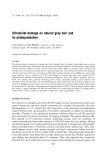
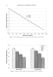
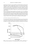
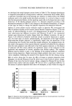
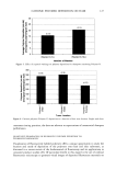
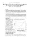
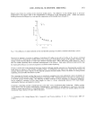
100 JOURNAL OF COSMETIC SCIENCE of the sun spectrum. It is inconceivable to limit the protection to absorption only. This would lead to a colossal storage of energy that would need to be dissipated by the cell. Such a process still remains unknown. The effects of sun exposure on the physiology of the epidermis have largely been reported in the literature. The well-known hyperkeratosis (25,26) has been measured i, vivo for the first time by confocal microscopy (Figure 5). This demonstrates the unique perfor- mance of this device in measuring noninvasively the thickness of the epidermal layers. Today, confocal microscopy remains the only technology providing rapid thickness measurements of the epidermal layers with a precision on the order of the micrometer. This avoids the need to perform painful biopsies on human volunteers. A 25% increase in stratum corneum thickness on each of the two subjects was recorded for a period of one month post-exposure. In this experiment, the epidermal hyperplasia of the epidermis i, vivo could not be determined. The only explanation for this is that such a measurement needs a marker formed by the bright signal of the basal keratinocytes that was lacking after exposure. Sun-exposed forearms led to unusual images through the confocal microscope. These unusual images were not observed before exposure and disappeared one month after exposure. These transient changes lasted for at least three weeks and consequently could not be attributed to a random phenomenon. The bright inclusions in the nuclei of numerous granular and spinous keratinocytes (Figure 6) can be interpreted as local condensations of chromatin. This would generate an index mismatch and consequently would lead to high reflections at the interface. Condensation of the chromatin seems to be in accordance with the histologic description of sunburn cells (27-29). Brightly spotted filaments in the intercellular spaces between keratinocytes (Figure 7) could correspond to aligned melanosomes migrating towards the surface via the den- drites of melanocytes (20,22). It has been stated that in normal conditions the dendritic epidermal cell lines could not be clearly identified with the i, vivo confocal microscope. This experiment has demonstrated, to the contrary, that only the dendrites but not the body of the melanocytes can be observed in exposed skin. The most striking transient observation turned out to be the total lack of melanosome caps in basal keratinocytes (Figure 8b) for a period of three weeks following sun expo- sure. A possible first explanation could be that photodegradation (30) or redox mecha- nisms (31) liberate the melanin contents of the melanosomes. Their geometry would thus not permit any further optical signal except for a diffuse absorption that is respon- sible for the tanned aspect of the upper layers. A second explanation could be related to the slow formation of melanosomes by the melanocytes, limiting both their increased migration towards the skin surface and the renewal of the reservoir in the basal kera- tinocytes (19-20). CONCLUSION The new phototype of CLSM dedicated to the exploration of human skin i, vivo provides major improvements when compared to previous systems. Sharper images were recorded with the blue laser line (488 nm), while the red line (647 nm) provided better pen-

IN VIVO CONFOCAL MICROSCOPY 101 etration into the dermis. The observation of unexpected details in epidermal cells, real-time tracking of blood cells and fast 3D reconstruction of the skin i, vivo, point to promising paradigms in skin research. These additional performances allowed for a definite explanation of the strong reflection of the basal keratinocytes: the melanosome caps acted as myriads of nanomirrors. A cascade of physiological events following sun exposure was recorded: a 25% increase in the stratum corneum thickness, a condensation of the chromatin in numerous kera- tinocytes, the suspected migration of the melanosomes via the dendrites of melanocytes, as well as the lack of melanosome caps in the basal keratinocytes. All of these events disappeared one month after exposure. It is now possible to observe that which was impossible before. I, vivo confocal micros- copy opens up new challenges in the understanding of skin optics and melanogenesis. ACKNOWLEDGMENT The authors wish to thank Daniel Good for his expert assistance in the correction of the manuscript. REFERENCES (9) (10) (11) (12) (13) (14) (1) M.J. Van Gemert, S. L. Jacques, H. J. Sterenborg, and W. Star, Skin optics, IEEE Trans. Biomed. Eng., 36, 1146-1154 (1989). (2) J. B. Dawson, D. J. Barker, D. Ellis, E. Grassam, J. A. Cotterill, G. W. Fisher, and J. W. Feather, A theoretical and experimental study of light absorption and scattering by in vivo skin, Phys. &led. Biol., 25,695-710 (1980). (3) P. H. Anderson and P. Bierring, Spectral reflectance of human skin in vivo, Photodermatol. Photoimmunol. Photomedo, 7, 5-12 (1990). (4) D. Contini, G. Zaccanti, F. Martelli, and A. Sassaroli, Models for photon migration and optical properties of biological tissues, Phys. Scr., T, 72, 76-82 (1997). (5) S. L. Jacques, Skin optics, Oregon &ledical Laser Center News (January 1998). (6) K.C. New, W.M. Petroll, A. Boyde, L. Martin, P. Corcuff J. L. L6v•que, M.A. Letup, H.D. Cavanagh, and J. V. Jester, In vivo imaging of human teeth and skin using real-time confocal micros- copy, Scanning, 13, 369-372 (1991). (7) P. Corcuff• C. Bertrand, and J. L. L•vSque, Morphometry of human epidermis in vivo by real-time confocal microscopy, Arch. Dermatol. Res., 285,475-481 (1993). (8) M. Rajadhyaksha, M. Grossman, D. Esterowitz, R. H. Webb, and R. R. Anderson, In vivo confocal scanning laser microscopy of human skin: Melanin provides strong contrast,J. Invest. Dermatol., 104, 946-952 (1995). P. Corcuff, G. Gonnord, G. E. Pi•rard, and J. L. L•vSque, In vivo confocal microscopy of human skin: A new design for cosmetology and dermatology, Scanning, 18, 351-355 (1996). B. R. Masters, G. Gonnord, and P. Corcuff Three-dimensional microscopic biopsy of in vivo human skin: A new technique based on a flexible confocal microscope, J. &licrosc., 185,329-338 (1997). S. Mallat, A theory for multiresolution signal decomposition: The wavelet representation, IEEE Trans. Pattern. Anal. Mach. Intdl., 11, 674-693 (1989). S. W. Zucker, Region growing: Childhood and adolescence, Cornput. Graphics Image Process., 5, 382- 399 (1976). T. B. Fitzpatrick, The validity and practicality of sun reactive skin type I through IV (editorial), Arch. DermatoL, 124, 869-871 (1988). D.T. Fewer, S.J. Hewlett, and E.M. McCabe, Laser sources in direct-view-scanning, tandem- scanning, or Nipkow-disk-scanning confocal microscopy, Applied Optics, 37, 380-385 (1998).
Purchased for the exclusive use of nofirst nolast (unknown)
From: SCC Media Library & Resource Center (library.scconline.org)