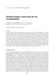
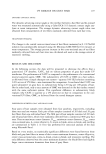
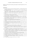
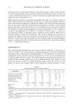
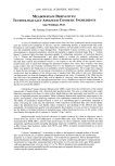
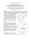
IN VIVO CONFOCAL MICROSCOPY 97 Figure 7. Sun exposure: migration of melanosomes. Two pictures showing the presence of bright spotted filaments in the intercellular spaces of the epidermal spinous layers. (a) Upper layers. (b) Lower layers. Subject B, T12. Image size: 168 x 168 pm. of 1.30 for a x40 objective lens certainly plays a role in increasing the axial and lateral resolution. Other lenses have been tested, in particular infinity-corrected objectives that introduced unexpected and unfortunate back reflections. Even though the laser power was limited to 2 mW at the objective lens, it provided a much larger light budget than the 1% of the light transmitted by the Nipkow disk of the TSM. There were also improvements made in the design of the contact stabilization device. By providing the axial displacement of the objective lens with a long-stroke piezoelectric translator with 0.1-pro precision, as opposed to moving the contact tip with a stepping motor of 1-pro step, a gain in precision was added to a gain in stability at the fixed skin surface. The Figure 8. Sun exposure: the lack of melanosome caps. (a) Before exposure, basal cells exhibit bright melanosome clusters. (b) After exposure, bright caps are almost missing a few are indicated by the white arrow. Subject B, T20. Image size: 168 x 168 pro.

98 JOURNAL OF COSMETIC SCIENCE resultant lateral stability was judged to be exceptional. Controlling the coarse approach of the confocal module to the skin surface with a motorized stage was found to be not only convenient but also very precise. The images of the keratinocytes exhibited unexpected details (Figure 2a). The plasma membranes were clearly depicted bright clusters and even granules in the cytoplasm could be observed. However, it is still impossible to unmistakably identify rare epider- mal cell lines such as melanocytes and Langerhans cells. The comparison of identical optical sections acquired with the three available wave- lengths did not produce the expected results (15). If the blue line gave sharper images than the red one, the difference remained relatively modest. On the other hand, even though the red line penetrates deeper into the skin, imaging with this wavelength seemed to be limited by the very strong scattering properties of the derreal network. The gain in depth could be estimated to be only 20 pro. A good compromise was provided by the intermediate laser line in the green region (568 nm) that allowed exploration down to the papillary dermis. However, images of the reticular dermis (150 pm in depth) could only be acquired with the red light. These images contain information not easily interpreted due to a lack of experience in the comprehension of images never before observed. The blood flow in the capillaries could be readily seen with the green laser. By zooming in on an individual capillary, time-resolved displacement of individual leukocytes could be followed. Direct videotape recording of live images remains the best mode of storage (16) since it does not have the recording time limitations of digital storage. It has been previously reported that melanin provides the contrast in the epidermal structures using in vivo confocal microscopy (7,8). Darker skin provides higher contrast. Thus, the bright bouquets and the crowns at the dertoo-epidermal interface (Figure 2b) are never seen in a plaque of vitiligo. Rarely seen in skin phototype I, they are observed in only 60% of phototype II and always in darker skins. The best contrast is obtained in Negroid skin from which increased papillomatosis and its big dermal papillae lead to spectacular images at the dermo-epidermal junction. The 3D reconstruction of the bright features from the basal cells (Figure 3a) allowed a definite association with the melanin caps (Figure 3b) covering the apical pole of the nucleus (17). Even though observed on vertical sections using a conventional transmis- sion light microscope, the melanin can easily be identified as absorbing clusters without employing any specific staining (Figure 9). The specific location of the melanin cap has been previously interpreted as a protection of the basal nuclei against injury from the sun (18). Absorption of light by melanin has been largely reported in the literature (19-21). Melanin absorption coefficients have been calculated. They increase rapidly at shorter wavelengths and reach a maximum in the UV region (5). The role of the melanin cap has been explained as a hat covering and protecting the nucleus to avoid DNA damage by UV rays (18). In the scanning electron microscope, the melanin cap consists of a cluster of capsule-shaped melanosomes (Figure 10a). Melanosomes are lisosome-like organelles containing tightly packed melanin proteins enclosed in a plasma membrane (22). They have a diameter of 200 nm and a length of 800 nm (Figure 10b). Their size is close to the Raleigh limit of the confocal microscope. Consequently, they have the potential to generate an optical signal in terms of reflection and/or multiple scattering
Purchased for the exclusive use of nofirst nolast (unknown)
From: SCC Media Library & Resource Center (library.scconline.org)