INHIBITION OF MATRIX METALLOPROTEINASES 231 Seoul) emitting wavelengths in the range of 340-450 nm. The radiation intensity was measured using a UV radiometer (EKO, Japan). The culture medium, DMEM, con- taining no serum, was added and incubated for 12 hours. The concentration of MMP-1 and -2 in the culture media was determined as described below. For the determination of NO in the cell body, cells were pretreated with nitric oxide sensor dye (1 pg/ml) (Clontech ApoAlert © nitric oxide/Annexin V dual sensor kit) and incubated for 30 minutes. The cells were UV-irradiated and incubated with the medium for 24 hours. Cells were washed twice with PBS and collected using trypsin-EDTA. The collection was centrifuged at 15,000 rpm for five minutes, and the supernatant was retained. This procedure was repeated twice. The pellet was resuspended with PBS. To analyze cells stained with NO sensor dye, a fluorescence-activated cell sorter (FACStarplus, Becton Dickinson) was used. ZYMOGRAPHY Zymography in sodium dodecyl sulphate-polyacrylamide gel electrophoresis (SDS- PAGE) containing 0.15% gelatin was performed according to the method of Demeule eta/. (16). The samples were mixed with SDS sample buffer in the absence of reducing agent, incubated at 37øC for 20 minutes, and electrophoresed on 10% polyacrylamide gels at 4øC. After electrophoresis, the gels were washed in 2.5% Triton X-100 for one hour to remove SDS and incubated for 12 hours at 37øC in 50 mM Tris-HC1, pH 7.6 0.15 NaC1 10 mM CaCI2 and 0.02% NAN3, and then stained with 0.1% Coomassie Brilliant Blue R250. DETERMINATION OF MMP-1 AND -2 BY ELISA The expression of MMP-1 and -2 was assayed by enzyme-linked immnosorbent assay (ELISA). HDFs (8 x 10³/well) were seeded into 96-well plates and cultured overnight. The culture media were replaced with DMEM containing SNP and/or AG. After incu- bation for 12 hours, the supernatants were transferred into a 96-well plate, and the coating buffer (Sa2CO 3 1.59%, NaHCO 3 2.93%, NaN 3 0.20%, MgCI 2 1.02%, pH 9.6) was added 1:1 (v/v) and incubated for 12 hours. The supernatants were removed and the coated well was washed with PBS-T three times, followed by blocking with 5% skim milk in PBS for one hour at 37øC. After washing three times with PBS containing 0.05% Tween 20 (PBST), 50 pl of 1/1000 diluted primary antibody, Ab-5 or Ab-3, in PBST was added into each well and incubated for 40 minutes. After washing the wells with PBST three times, 50 pl of 1/1000 diluted secondary Ab and anti-mouse IgG conjugated with alkaline phosphatase in PBST was added and incubated for 40 minutes. After washing five times with PBST, 100 pl of 1 mg/ml pNPP (p-nitrophenyl phos- phate) in diethanolamine buffer was added. The optical density was measured at 405 nm after 30 minutes. The cytotoxicity of the supplemented chemicals was measured by a 3-(4,5-cimethylthiazol-2-yl)-2,5-diphenyl tetrazolium bromide (MTT) assay. RT-PCR-ELISA Total RNA was extracted from cultured cells using the Promega RNAgent © total RNA isolation system. PCR amplification was carried out using 5' biotinylated primers

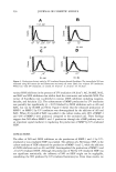
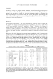
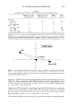
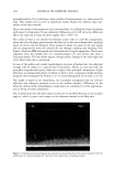
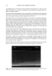
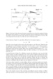
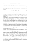
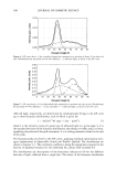
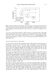
232 JOURNAL OF COSMETIC SCIENCE (sense) to generate biotinylated PCR products detectable by digoxygenin-labeled probes in an immunoenzymatic assay (ELISA) method (17). First, cDNA was mixed with 10x buffer, 10 mM dNTPs, 1 U Taq DNA polymerase (Bioneer, Korea), and iNOS primers in a final volume of 50 lal. The primer sequences and relative predicted PCR product sizes are given below: Human iNOS (sense) 5'-AGTTTCTGGCAGCAACGG-3' Human iNOS (anti-sense) 5'-TTAAGTTCTGTGCCGGCAG-3' A sample containing all reaction reagents except cDNA was used as PCR negative control in any amplification. The mixtures were incubated for the indicated cycles (predenaturation 5 min at 95øC denaturation 50 sec at 95øC annealing 20 sec at 56øC extension 20 sec at 72øC) in a GeneAmp PCR System 2400 (Perkin-Elmer). The correct size of all PCR products was confirmed by comparing with a DNA standard on agarose gel. After a given cycle of PCR, the amount of amplified cDNA was determined by the ELISA method. First, microplates (Maxisorp Nunc) were coated with 50 lag/ml of avidin (Sigma) in coating buffer (CB 15 mM Na2CO3, pH 9.6) and incubated for two hours at 37øC. After incubation, free sites were saturated with 2% blocking solution (Roche, Germany) in CB. Biotinylated PCR products diluted in PBS containing 3% bovine serum albumin (PBSB) were distributed onto microplates (100 lal per well) and incu- bated for one hour at room temperature. After incubation, the microplates were washed three times with PBST. Amplified cDNA was denatured using 0.25 M NaOH at room temperature for ten minutes. Following the washing, 100 lal per well of 10 pmol/ml digoxygenin-labeled probes in hybridization buffer [6.25x SSC, 0.625% blocking re- agent (Roche), 0.125% Tween 20, and 0.5 M NaH2PO 4 (pH 6.5)] were added and incubated at 42øC for two hours. Anti-digoxygenin AP-conjugated antibody (Sigma) was added (1:3000 in PBSB) and incubated for one hour at 37øC. The reaction was developed by nitrophenyl phosphate (pNPP 1M diethanolamine buffer, pH 9.6). The amount of amplified product was measured for optical density at 405 nm (OD 405) using a microplate reader. STATISTICAL ANALYSIS Results were presented as means + standard error (SE). Experimental results were statistically analyzed by using Student's t-test (SigmaPlot 2000). P values 0.05 were regarded as indicating significant differences. RESULTS AND DISCUSSION EFFECT OF UV ON THE PRODUCTION OF MMPS The immunoreactive MMP-1 and -2 in the culture medium of HDFs were measured using anti-MMP-1 and -2 monoclonal antibodies, respectively. Treatment of HDFs with UV radiation enhanced the production of MMP-1 by twofold and MMP-2 by threefold in a dose-related manner (Figure 1), confirming the previous results (1-5). We also confirmed that the gelatinase activities were proportionally increased by the UV irra- diation of HDFs using gelatin zymography (data not shown). The production of MMPs
Purchased for the exclusive use of nofirst nolast (unknown)
From: SCC Media Library & Resource Center (library.scconline.org)