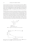
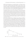
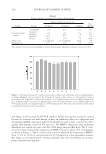
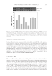
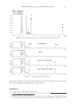
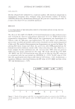
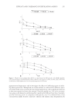
ANTI-WRINKLE ACTIVITY OF P. STROBILACEA 221 Figure 6. Photographic assessment of wrinkles in the crow’s feet area of (a) the test group and (b) the pla- cebo group by CCD camera after the use of the formulation containing P. strobilacea fruit extract for 12 weeks. Figure 5. Weekly comparison of R3 (average roughness) values measured by the Skin-Visiometer SV 600 (Courage & Khazaka, Germany) in the crow’s feet area after the use of the formulation containing P. strobila- cea fruit extract for 12 weeks (*p 0.05). the average roughness, is the arithmetic average of the different segment roughnesses. Inherent in their defi nitions, R3 is the most adequate parameter for studying. The test formulation also showed a signifi cant improvement in the average difference in roughness (ΔR3, −0.02 ± 0.06) than did the placebo formulation (ΔR3, 0.01 ± 0.05) 12 weeks after the treatment (Figure 5). In the clinical study of measurements using visual evaluation and image analysis, the test cream showed a signifi cantly different effect (p 0.05) from that of the placebo (Figure 6). CONCLUSIONS In this study, in order to investigate the potential of P. strobilacea fruit extract as an active ingredient for wrinkle-care cosmetics, we measured their free-radical scavenging activity, elastase inhibitory activity, the expression of MMP-1 in vitro, and type I collagen synthe- sis in normal human fi broblast cells. P. strobilacea fruit extracts (SC50 5 μg/ml) showed very high free-radical scavenging activity compared to BHT (SC50 = 25.4 μg/ml) as a positive control. Elastase inhibitory activity of P. strobilacea fruit extract (IC50 = 37.9 μg/ml)

JOURNAL OF COSMETIC SCIENCE 222 was over two times higher than that of oleanolic acid (IC50 = 83.8 μg/ml). P. strobilacea fruit extract reduced the expression of MMP-1 mRNA (up to about 70% at 50 μg/ml). Ellagic acid was isolated as a main component and identifi ed using spectroscopic analysis. In the MMP-1 mRNA expression assay on human fi broblast cells stimulated by UVA radiation, ellagic acid slightly reduced the expression of MMP-1 mRNA about 15% at 1 μg/ml while P. strobilacea fruit extract reduced it about 70% at 50 μg/ml. P. strobilacea fruit extract and ellagic acid increased the expression of type I collagen mRNA in a dose- dependent manner (up to 37% and 41% at 20 μg/ml and 1.0 μg/ml, respectively), compa- rable to that of ascorbic acid (up to 39% at 500 μM). P. strobilacea fruit extract showed no cytotoxicity up to the effective concentration for anti-wrinkle activity. In this clinical study, the test and placebo formulation (the test cream contained 0.2% P. strobilacea fruit extract and the placebo did not) were compared. The visual evaluation and image analysis used in this study showed a statistically signifi cant difference (p 0.05) between the ef- fects of the tested formulation and the placebo 12 weeks after the treatment. All of these results suggest that Platycarya strobilacea fruit extract could be used as an active ingredi- ent for new anti-wrinkle cosmetics. ACKNOWLEDGMENTS This work was supported by a grant from the Korean Ministry of Commerce, Industry, and Energy, entitled “The development of cosmeceutical ingredients from natural plants like Nymphaea tetragona of Jeju plant” (Subject Number 2006-10027299), as part of a regional specialization technique development project within a regional industry promo- tional project of the Ministry of Commerce, Industry, and Energy. REFERENCES (1) B. A. Gilchrest, M. Garmyn, and M. Yaar, Aging and photoaging affect gene expression in cultured human keratinocytes, Arch. Dermatol., 130, 82–86 (1994). (2) J. H. Chung and H. C. Eun, Angiogenesis in skin aging and photoaging, J. Dermatol., 34, 593–600 (2007). (3) B. A. Gilchrest, Skin aging and photoaging: An overview, J. Am. Acad. Dermatol., 21, 610–613 (1989). (4) A. Perrin, E. Bauza, C. D. Farra, and N. Domloge, Stimulating effect of collagen-like peptide on the extracellular matrix of human skin: Histological studies, Int. J. Tissue React., 26, 97–104 (2004). (5) G. J. Fisher, The pathophysiology of photoaging of the skin, Cutis, 75, 5–8 (2005). (6) J. H. Chung, S. H. Youn, O. S. Kwon, H. C. Eun, K. H. Kim, K. C. Park, K. H. Cho, and J. I. Youn, Enhanced proliferation and collagen synthesis of human dermal fi broblasts in chronically photodam- aged skin, Photodermatol. Photoimmunol. Photomed., 12, 84–89 (1996). (7) H. Birkedal-Hansen, W. G. Moore, M. K. Bodden, L. J. Windsor, B. Birkedal-Hansen, A. DeCarlo, and J. A. Engler, Matrix metalloproteinases: A review, Crit. Rev. Oral Biol. Med., 4, 197–250 (1993). (8) P. Brenneisen, J. Oh, M. Wlaschek, J. Wenk, K. Briviba, C. Hommel, G. Herrmann, H. Sies, and K. Scharffetter-Kochanek, Ultraviolet B wavelength dependence for the regulation of two major matrix- metalloproteinases and their inhibitor TIMP-1 in human dermal fi broblasts, Photochem. Photobiol., 64, 877–885 (1996). (9) Y. Kawaguchi, H. Tanaka, and T. Okada, The effects of ultraviolet A and reactive oxygen species on the mRNA expression of 72-kDa type IV collagenase and its tissue inhibitor in cultured human dermal fi - broblasts, Arch. Dermatol. Res., 288, 39–44 (1995). (10) H. Watanabe, T. Shimizu, J. Nishihira, R. Abe, T. Nakayama, M. Taniguchi, H. Sabe, T. Ishibashi, and H. Shimizu, Ultraviolet A-induced production of matrix metalloproteinase-1 is mediated by macrophage migration inhibitory factor (MIF) in human dermal fi broblasts, J. Biol. Chem., 279, 1676–1683 (2004).
Purchased for the exclusive use of nofirst nolast (unknown)
From: SCC Media Library & Resource Center (library.scconline.org)