J. Cosmet. Sci., 63, 297–302 (September/October 2012) 297 Aloe-emodin inhibits proliferation of adult human keratinocytes in vitro DUSAN POPADIC, EMINA SAVIC, ZORICA RAMIC, VLADIMIR DJORDJEVIC, VLADIMIR TRAJKOVIC, LJILJANA MEDENICA, and SVETLANA POPADIC, Institute of Microbiology and Immunology (D.O., E.S., Z.R.,V.T.), Department of Dermatovenereology (L.M., S.P.), and Department of Otorhinolaryngology and Maxillofacial Surgery (V.D.), Faculty of Medicine, University of Belgrade, Belgrade, Serbia. Accepted for publication January 17, 2012. Synopsis Aloe-emodin (AE) is a plant-derived hydroxyanthraquinone with several biological activities. It is present in a variety of skin-conditioning agents containing aloe extracts, but its infl uence on keratinocyte growth was not examined so far. We investigated the infl uence of AE on human keratinocyte proliferation and apoptosis in vitro. AE signifi cantly inhibited proliferation of cultivated human keratinocytes at 5 μM concentration, as revealed by incorporation of radioactive thymidine. The antiproliferative effect of AE was accompanied with induction of apoptosis, but not necrosis, as demonstrated by fl ow cytometric analysis and lactate dehy- drogenase release assay. Based on the half maximal inhibitory concentration values, we demonstrated that AE may impair proliferation of keratinocytes at concentrations far below the industry standards for com- mercial products containing aloe extracts. Therefore, further research of AE effects on the human skin and proper labeling of products are necessary for maximizing benefi ts from aloe extracts and to avoid undesired responses. INTRODUCTION Since ancient times, Aloe vera has been used to help wound and burn healing. Several re- ports emphasize the stimulatory effect of whole extracts or components from aloe on skin cell proliferation and collagen synthesis (1). By the virtue of these effects, aloe is currently used in a variety of commercial products, including sunscreens, cosmetics, and lotions. Aloe crude extracts typically present in topical formulations contain a variety of active substances differing considerably between manufacturers and from batch to batch (2). Moreover, the functional relationship between the various compounds and their net effects on human cells has not been completely established. Address all correspondence to Dusan Popadic at dpopadic@med.bg.ac.rs. Dusan Popadic and Emina Savic contributed equally to this work.

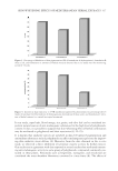
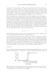
JOURNAL OF COSMETIC SCIENCE 298 Aloe-emodin (AE), a hydroxyanthraquinone from Aloe vera leaves has been recognized for a long time as a potent laxative, while in the last decade, several other biological activities are reported including its antiproliferative effect (3). On the contrary, there are data indi- cating that AE stimulates growth and increases DNA synthesis in some primary cell cultures (4). Although it is present in topical formulations that are applied to the skin, the infl uence of AE on normal keratinocytes has not been examined so far. In this study, we investigated the effect of AE on primary adult keratinocyte monolayer cultures. Our data indicates that AE signifi cantly reduces proliferation capacity of the keratinocytes cultivated in vitro at doses as low as 5 μM, and this effect can be attributed to the induc- tion of apoptosis. MATERIAL AND METHODS CELL CULTURE AND REAGENTS Skin samples were obtained from 10 subjects undergoing cosmetic surgery, who all gave the informed consent, and processed as described previously (5). Cells were used for experi- ments after the third or fourth passage. AE was dissolved in dimethylsulfoxide (DMSO) (both from Sigma, St. Louis, MO). Control cell cultures contained same amount of DMSO as cultures with the 5 μM concentration of AE used in the particular experiment. CELL PROLIFERATION AND LACTATE DEHYDROGENASE (LDH) RELEASE ASSAY Keratinocytes were cultivated in 96-well plates (3 × 10³ cells/well) for 48 h, then washed and cultivated for an additional 72 h in fresh medium containing 1.25, 2.5, or 5 μM AE or DMSO as a control. During the last 24 h of incubation, keratinocytes were pulsed with 1 μCi of [3H]thymidine per well and proliferation was determined as described previously (5). The half maximal inhibitory concentration (IC50) values for the inhibition of prolifera- tion were calculated using Calcusyn software (Biosoft, Cambridge, UK) (5). LDH release assay was employed to assess cell necrosis. Keratinocytes were cultivated under the same condi- tions as for the proliferation assay and LDH assay was performed as previously described (6). DETERMINATION OF APOPTOSIS For the assessment of apoptosis, keratinocytes were cultivated in 24-well plates (3 × 10⁴/ well) for 48 h, washed and incubated for an additional 6 h in fresh medium containing 5 μM of AE or DMSO as a control. Following trypsinization, the cells were stained with fl uorescein diacetate (FDA) and trypan blue (TB), and analyzed as described previously (5). STATISTICAL ANALYSIS Statistical signifi cance of the differences in keratinocyte proliferation in the group of 10 donors was determined by the analysis of variance (ANOVA) for repeated measures.
Purchased for the exclusive use of nofirst nolast (unknown)
From: SCC Media Library & Resource Center (library.scconline.org)