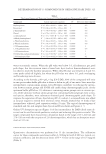
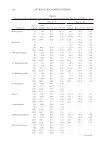
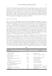
SKIN-WHITENING EFFECTS OF MEDITERRANEAN HERBAL EXTRACTS 313 by Folin–Ciocalteu method (20): 24% in Capparis spinosa, 25% in Citrus sinensis, 22% in Oryza sativa, 27% in Olea europaea extract, and 26% in their mixture. Mushroom tyrosinase, L -DOPA, and other reagents were purchase from Sigma-Aldrich (Milan, Italy). Topical application of active compounds was performed by oil-in-water emulsions, adding skin-whitening ingredients, as reported in Table I. The emulsions were prepared by slowly adding the aqueous phase to the oily phase and a blend of surfac- tants under continuous agitation both phases were kept at 70°C. This mixture was stirred until it was cool, thus forming emulsion formulations. IN VITRO EVALUATION OF ANTI-TYROSINASE ACTIVITY The in vitro tyrosinase inhibitory activity (IA) was determined by the dopachrome method. Herbal extracts and their combination were added at a concentration of 50 μg/ml to L-DOPA solution (10 mM, dissolved in a 5 mM acetate buffer, pH 5). Subsequently, ty- rosinase enzyme was added to each solution to a fi nal concentration of 4 U/ml. The amount of dopachrome in the reaction mixture was measured after incubation at 38°C for 60 min at 475 nm by UV-Vis spectrophotometer (UV-1700 PharmaSpec SHIMADZU Milan, Italy). Kojic acid and hydroquinone, at the concentration of 50 μg/ml, were used as reference and a blank reaction was conducted without a sample. All measurements were performed in triplicate. The activity of each sample was expressed as the IA using equation 1: u100 blank sample blank OD OD IA (%) OD (1) where ODblank and ODsample are the optical density values of the blank and the solutions containing active ingredients, respectively. IN VIVO EVALUATION OF SKIN-WHITENING ACTIVITY In vivo experiments were performed on 15 healthy volunteers (females/males 10:4) of skin types II and III, aged 30–45 years. Volunteers were recruited after medical screening in- cluding the fi lling of a health questionnaire followed by physical examination of the ap- plication sites. Subjects exhibiting such features as sun-burn, suntan, burn marks, or any other active lesions, which might interfere with evaluation, were excluded from the study. After they were fully informed of the nature of the study, substances, and procedures involved, Table I Composition of Oil-in-Water Emulsions Used as Topical Skin-Whitening Formulations Oil-in-water emulsion Oil phase (PPG-15 stearyl ether-8 g, isohexadecane\PPG-15 stearyl ether-4 g) Aqueous phase (Distilled water-73.7 g) Surfactants and structurizing agents (Steareth 2–3.5 g, steareth 2.1–2.5 g, stearic acid-2.5 g, cetylstearylic acid-2.1 g, xanthan gum-0.3 g, ethylene glycolphenyl undecylether p-hydroxybenzoate-0.4 g) Formulation A Hydroquinone-3 g Formulation B Kojic acid-3 g Formulation C Mixture of herbal extracts-3 g

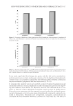
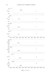
JOURNAL OF COSMETIC SCIENCE 314 they gave their written consent. Two research assistants were responsible for all recruitment and data collection. The in vivo experiments were carried out on the volar forearms of each volunteer, and each subject was rested for 15 min before the experiments and room conditions were set at 22 ± 2 °C and 40–50% relative humidity. Four sites on the ventral surface of each forearm were defi ned using a circular template (1 cm2) and demar- cated with permanent ink. During the fi rst week of experiments, skin tanning was in- duced in each site by exposition of a lamp that simulate sunlight (Helios Italquartz srl, Milan, Italia), which emitted in a range of 300–400 nm (6.5 mW cm−2), for 2–8 min depending on the minimal erythema dose (MED) of each subject. The initial melanin value and the melanin content obtained during the monitoring period were performed by refl ectance visible spectrophotometer X-Rite model 968 (X-Rite Inc., Grandville, MI) as previously reported (21). The variation of skin melanin values was monitored for a total period of 4 weeks. To avoid induced and interfering skin erythema events, exposition to a lamp was not conducted on the third and the sixth day of the fi rst week. For each forearm, three skin sites were treated with tested formulations and the one not treated was consid- ered as control. Tested formulations were applied after each lamp treatment for the fi rst week and once daily in the second, third, and fourth week of the study. To evaluate the time course of skin pigmentation, melanin index (MI) baseline values were subtracted from the MI values to calculate mean MI (ΔMI) values. For each site, plotting ΔMI vs. time, the area under the curve was computed using the trapezoidal rule to obtain area under curve (AUC) dimensionless index values directly related to the degree of skin pig- mentation. From the AUC values obtained, the skin-whitening effect of each formulation was expressed as the inhibition of skin pigmentation (PI) using the following equation: u100 AUC AUC PI (%) AUC C S C (2) where AUCC is the AUC value of no treated skin site and AUCS is the AUC value ob- tained from treated sites. IN VIVO EVALUATION OF PHOTOSENSITIZING EFFECT OF SKIN-WHITENING AGENTS The skin tolerance of skin-whitening agents was investigated by an in vivo model previ- ously reported (21). After a rest period of 6 months, the same subjects participating in the previous study were enrolled to evaluate if skin-whitening application had increased the skin sensitivity to UVB irradiation. For each subject, four skin test sites were defi ned on the ventral surface of each forearm. Three sites were treated with formulations once daily for 4 consecutive weeks and one site was used as control (no topical treatment). At the end of the fourth week, all sites were exposed to UVB irradiation dose, corresponding to the MED by using the lamp previously described to simulate sunlight. The induced ery- thema was measured, after 24 h from the skin site exposure, by refl ectance spectropho- tometry and the registered photosensitivity was expressed as a percentage calculated from erythema index values using equation 3: u100 EI EI Photosensitivity (%) EI T C C (3) where EIC is the erythema index of control sites and EIT is the erythema index of sites treated with formulations.
Purchased for the exclusive use of nofirst nolast (unknown)
From: SCC Media Library & Resource Center (library.scconline.org)