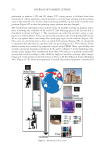
The SCC’s new Media Library and Resource Center is a digital platform poised to be the industry’s go-to resource for the latest scientific research, education and information. Content includes: x Digital issues of the Journal of Cosmetic Science x Archived SCC Webinars x Videos/slide-syncs of scientific presentations delivered at the Annual Meeting x and so much more. Start exploring today! www.scconline.org/library www.scconline.org/library

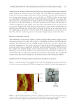
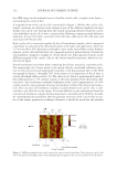
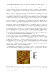
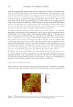
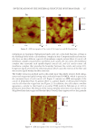
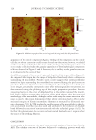
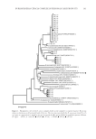
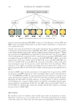
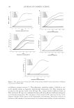
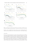
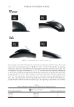
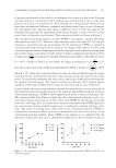
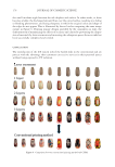
J. Cosmet. Sci., 71, 117–131 (May/June 2020) 117 Investigation of the Internal Structure of Human Hair with Atomic Force Microscopy ROGER L. MCMULLEN and GUOJIN ZHANG, Ashland Specialty Ingredients G.P., Bridgewater, New Jersey 08807 (R.L.M., G.Z.) Accepted for publication February 6, 2020. S ynopsis The internal ultrafi ne structure of human hair was explored with atomic force microscopy (AFM). Cross sections of hair were prepared by a proprietary technique that provided a smooth surface for effective imaging in contact-mode AFM. Investigations of virgin hair revealed structural details of cortical and cuticle cells consistent with previous transmission electron microscopy (TEM) studies, in addition to the identifi cation of a boundary region surrounding macrofi brils of the cortex. The effects of bleaching and solvent extraction on the internal structure of hair were also investigated. In the cuticle cell, bleaching causes the most damage to the endocuticle and cell membrane complex, evident by erosion of these components. Similarly, bleaching results in crevices, cracks, and asperities in the cortex of hair. In addition, the cortical cell membrane complex appears compromised along with either lipid or protein structures at the outer boundaries of macrofi brils. In delipidated hair, most structural components of the fi ber appear intact with the exception of an overall swollen nature of the various morphological components. INTRODUCTION The ultrafi ne structure of human hair has captured the attention of scientists for decades. Most of what we currently know about hair morphology comes from a plethora of studies of thin sections of hair using transmission electron microscopy (TEM) (1–5). Despite advances made in our understanding of the hair structure, there still remain a number of unanswered questions about the physicochemical properties of hair. The advent of atomic force microscopy (AFM) techniques gave researchers an alternative route to explore the nanomechanical, nanotribological, and nanostructural properties of the hair fi ber. AFM studies of human hair started to appear in the literature in the late 1990s to early 2000s and mostly focused on the quantitative analysis of three-dimensional topographi- cal images of the hair surface (6–8). A key advantage to using AFM as an investigative technique of the morphological properties of the hair fi ber surface is that there is no need to coat the specimen with metal (e.g., gold and platinum) and studies can be conducted in ambient conditions (and even in solution) rather than vacuum (9). A study of cuticle Address all correspondence to rmcmullen@ashland.com. Current affi liation: Guojin Zhang, L’Oréal, Clark, New Jersey 07066.
Purchased for the exclusive use of nofirst nolast (unknown)
From: SCC Media Library & Resource Center (library.scconline.org)