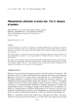
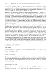
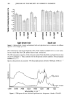
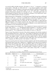
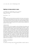
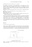
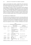
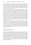
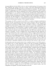
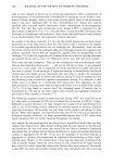
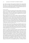
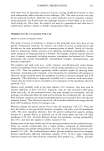
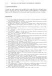
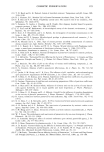
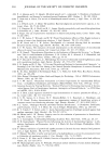
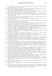
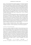
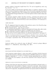
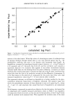
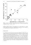
186 JOURNAL OF THE SOCIETY OF COSMETIC CHEMISTS v• 4,5 • 4 e- 3,5 0'• 3 .c= 2,5 _.m 2 E 1,5 0 1 =__ 0,5 ß -• O. : : : : : I : : : : : : m '-- o m '-- o • • • -- _ • •) • -- _ light-brown hair black hair Figure 2. Melanin yields of intact and irradiated black and light-brown hair in relation to the different segments of the sunlight spectrum. The transmission spectrum (diamond cell) of the melanin samples led to a more satis- factory result than the KBr pellets (better band resolution). The IR transmission spectra of the melanin isolated from the black and light-brown hair are given in Figure 3. They conform well to the spectra of both synthetic (4) and natural melanin (12,17). --1 Spectral characteristics ofeumelani,. The broad absorptions between 3600 and 2500 cm 2 b[c]ck • • [ighf-brow i I i I i j 4000 3680 2960 2460 1920 1400 880 360 Wovenumber in crn -• Figure 3. FTIR spectra of intact black and light-brown hair. Assignment of bands of Table III.

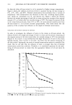
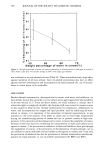
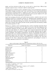
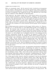
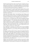
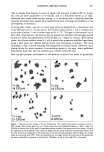
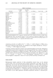
HAIR MELANIN 187 are caused by OH- and NH-vibrations. The peak at 1710 cm- • is assigned to a carbonyl group (peak 1, Table III). At 1620-1630 cm-•, ring vibration appears, which might be overlapped by an NH-vibration. Ring structures typical for melanin involve pyrrone, y-thiopyrrone, indole, and quinones (peak 2) (12). The small peak 3 indicates the presence of heterocyclic moieties. At 1220 and 1157 cm-• two peaks (peak 4 and 5) of similar height are observed they were identified by Boudier (4) as characteristic vibra- tions of the dihydroxyindole structure in melanin. Spectral characteristics ofpheomelanin. In the IR spectrum of light-brown hair an additional vibration at 1082 cm- • is found. It is possible that it arises from the presence of sulfur --1 moieties in the pheomelanin that produce vibrations in the region 1070-1110 cm (S-aromatic compounds, peak 6 in Table III) (18). This absorption is, apart from the SEM photographs of the melanin granules and color values of the nonirradiated hair (Table I), a third point of evidence indicating the difference in the overall structures of the pigments in the investigated hair samples. IR spectra of melanin from irradiated hair. The comparison of spectra is made only for the region 1900-550 cm- • i e. the absorption bands of melanin: C = O (peak 1), quinone (peak 2), heterocyclics (peak 3), dihydroxyindole (peaks 4, 5), and S-aromatic com- pounds (peak 6). The corresponding wave numbers are given in Table III. Figures 4a and 4b provide the spectra of the melanin pigments derived from irradiated and nonirradiated black hair, respectively. The changes in absorption of the black hair melanin are evident only for hair that was irradiated with visible light (Figure 4b) peaks 4 and 5 are slightly visible. A similar but much smaller effect is seen for samples irradiated with UV-A light. It appears thus that visible light, and to much lesser extent UV-A, degrades the DHI structure of the melanin in black hair. Figures 5a and 5b provide the spectra of melanin pigments derived from irradiated and nonirradiated light-brown hair, respectively. Comparison of the spectra of the irradiated light-brown hair pigments shows clearly the different effects of the segments of sun- light. UV-B, UV-A, visible light, IR, and global irradiation lead to a destruction of the absorption bands of C = O (peak 1) and quinone (peak 2) (Figures 5a and 5b). Visible light, UV-A, and global irradiation show a particularly strong effect. In addition, Table III IR Vibrations of the Melanin Polymer and the Corresponding Functional Groups Peak number Region (cm- 1) Description of peak Functional groups 3600-2500 Broad absorption OH, NH 1710 Shoulder C = O, COOH (9) 1628 Significant peak quinone, pyrrole, thiopyrrole (9) 1420 Small peak heterocycles 1220 Two peaks with similar height dihydroxi-5,6-indol 1157 polymer (4) 1082 Significant peak ring vibration with C-S- interaction (18) i In black hair. 2 Additionally in light-brown hair.
Purchased for the exclusive use of nofirst nolast (unknown)
From: SCC Media Library & Resource Center (library.scconline.org)