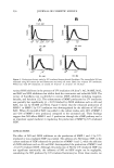
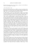
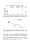
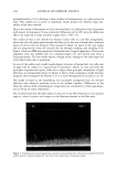
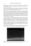
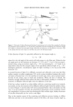
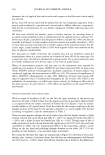
HAIR STRAND TEST 269 2 4 8 Figure 2. Growth ofM. globosa CBS 7966 after 18-day incubation hair from ten volunteers pre-incubated in andidandruff shampoo (A) (growth 1/10 Co = control). markedly lower concentration compared with that of climbazole, but may also be a specific feature of the substance, which is revealed by the hair strand test. Previous in vitro studies have shown hydroxypyridones to be highly effective against Malassezia spp., but data on their bioavailability in/on human hair are not yet available this also applies to the other agents tested. The polidocanol concentrations used should actually have had an inhibitory effect (18), but probably this substance, too, does not bind to human hair, so that such an effect could not be demonstrated. Tests using higher concentrations might be reasonable. CONCLUSION In summary, the in vitro hair strand test was found to be an interesting and reliable new test model for evaluation of the antifungal activity of antidandruff preparations, espe- cially with regard to a possible depot effect. Climbazole proved to be effective. With all other agents, no bioavailability from the hair was found, possibly because of the low concentrations used. Other substances (e.g., zinc pyrithione, ketoconazole, selenium disulfide, tar) are currently tested with the new system. The current test model does not primarily assess binding of antimycotics to scalp keratin. Active ingredients might rediffuse from the compartment of the hair to influence the growth of Malassezia yeasts on the sebum-rich scalp surface. Supplementary examination of the hair samples by GC-MS analysis would substantiate the validity of the test system. The hair strand test could also be performed ex vivo with hair samples from volunteers who regularly use

270 JOURNAL OF COSMETIC SCIENCE antidandruff preparations. In addition, the effects of repeated washing with or without active ingredients (wash-out kinetics, saturation effects) could also be evaluated. REFERENCES (1) E. Gu•ho, T. Boekhout, H. R. Ashbee, et aL, The role of Malassezia species in the ecology of human skin and as pathogens, Med. MycoL, 36 (suppl. 1), 220-229 (1998). (2) G. Midgley, The lipophilic yeasts: State of the art and prospects, Med. Mycol., 38 (suppl. 1), 9-16 (2000). (3) E. Gu•ho, G. Midgley, and .J. Gulllot, The genus AJa/assezJa with description of four new species, Antonie van Leeuwenhoek, 69, 337-355 (1996). (4) J. Guillot, E. Gu•ho, M. Lesourd, et al., Identification of Malassezia species: A practical approach. J. Mycol. Mgd., 6, 103-110 (1996). (5) R.J. Hay and C. Graham-Brown, Dandruff and seborrheic dermatitis: Causes and management, Clin. Exp. Derm., 22, 3-6 (1997). (6) C. Pi•rard-Franchimont, J. F. Hermanns, H. Degreef, and G. E. Pi•rard, From axioms to new insights into dandruff, Dermatology, 200, 93-98 (2000). (7) R.D. Aron-Bruneti•re, D. Dompmartin-Pernot, and E. Drouhet, Treatment of pityriasis capiris (dandruff) with econazole nitrate, Acta Derm. Venereol., 57, 77-80 (1977). (8) L. C. Barber, The aetiology of dandruff. PR No. 8. Pityrosporum ovale/amphotericin B dandruffaetiology study (DA-125). Internal Report. Procter & Gamble, Cincinnati, 1977. (9) M.M. Carr, D. Pryce, and F.A. Ive, Treatment of seborrheic dermatitis of the scalp with 2% ketoconazole, Br. J. Dermatol., 116, 213-216 (1986). (10) S. Shuster, The aetiology of dandruff and mode of action of therapeutic agents, Br. J. DermatoL, 111, 235-242 (1984). (11) M. C. Y. Heng, C. L. Henderson, D.C. Barker, et aL, Correlation of Pityrosporum ovale density with clinical severity of seborrhoeic dermatitis assessed by a simplified technique, J. Am. Acad. DermatoL, 23, 82-86 (1990). (12) R. W. Van der Wyck and F. C. Roia, The relationship between dandruff and the microbial flora of the human scalp, J. Soc. Cosmet. Chem., 15, 761-768 (1964). (13) R. W. Van der Wyk and K. E. Hechemy, A comparison of the bacterial and yeast flora of the human scalp and their effect upon dandruff production,.]. Soc. Cosmet. Chem., 18, 629-639 (1967). (14) I.M. Bergbrant and J. Faergemann, Seborrhoeic dermatitis and Pityrosporum ova/e: A cultural and immunological study, Acta Derre. Venereol., 69, 332-335 (1989). (15) D.C. Clift, H. J. Dodd, J. D. T. Kirby, et aL, Seborrhoeic dermatitis and malignancy. An investigation of the skin flora, Acta Derre. VenereoL, 68, 48-52 (1988). (16) N.J. Van Abbe, The investigation of dandruff, J. Soc. Cosmet. Chem., 15, 609-630 (1964). (17) K.J. McGinley, L.J. Leyden, R. R. Marpies, et aL, Quantitative microbiology of the scalp in non- dandruff, dandruff, and seborrheic dermatitis, J. Invest. DermatoL, 64, 401•405 (1975). (18) P. Mayser and K. Grtinder, Growth inhibition of Malassezia species by pharmacological concentrations of polidocanol, Mycoses, 38, 23-27 (1995).
Purchased for the exclusive use of nofirst nolast (unknown)
From: SCC Media Library & Resource Center (library.scconline.org)