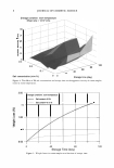
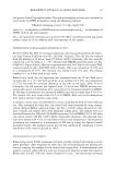
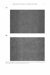
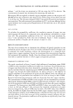
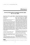
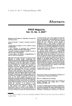
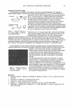
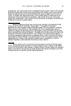
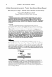
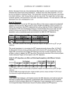
18 JOURNAL OF COSMETIC SCIENCE (a) Figure 1. Microphotographs of the original emulsion. (a) Without crossed polarizers. (b) With crossed polarizers. Magnification: (a) 400x (b) l00x.

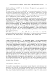
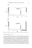
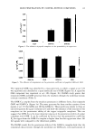
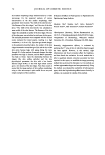
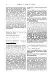
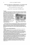
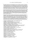
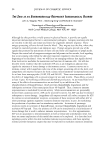
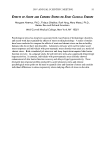
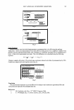
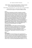
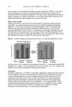
125 • e 100 - ,, 0 '- 75 - CD i 50 25 0 0 CHANGES DURING EVAPORATION OF EMULSION 1 2 Diameter, microns 3 19 4 Figure 2. The number size distribution of the original emulsion (6) and after evaporation to 60% of 40% of the initial weight (■). crystal, but the particles are too small to provide an answer. However, when one realizes that the emulsion remains at this stage are the essential element in order to understand the relation between the emulsion components and its action on the skin, it is apparent that additional information is obviously of essence. Some of this informational needed is offered by the results of the centrifugation (Figure 7). The bottom layer emulsion is now increased to 73% of the emulsion Figure 3. Original emulsion after centrifugation.
Purchased for the exclusive use of nofirst nolast (unknown)
From: SCC Media Library & Resource Center (library.scconline.org)