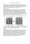
62 JOURNAL OF COSMETIC SCIENCE Aldrich) in water. The solution was diluted in a calibrated flask. The complex of GSH with Cu was molar ratio 2:1 and 1:1. • A buffer solution (pH 7.4) was prepared by dissolving potassium phosphate (POCH, Gliwice, Poland), and its pH was adjusted to 7.4 by addition of di-sodium hydrogen phosphate dodecahydrate (POCH, Gliwice, Poland). The obtained solution was di luted to 1000 ml with demineralized water. • A 0.1 % biscyclohexanon-oxalyldihydrazone (cuprizon) (Fluka, Buchs, Switzerland) solution was prepared by dissolving 200 mg of cuprizon in 40 ml of hot 50% ethanol. This solution was diluted with ethanol to 200 ml. • A buffer solution (pH 10.0) was prepared by dissolving ammonium chloride (POCH, Gliwice, Poland) and was adjusted to 10.0 by addition of ammonium (POCH, Gli wice, Poland). The obtained solution was diluted to 1000 ml with demineralized water. • Trifluoroacetic acid (TFA) solution (0.15%) (Fluka, Buchs, Switzerland) was prepared by dissolving an appropriate amount in distilled water. The solution was diluted in a calibrated flask. • Components of the model emulsion: 8% glyceryl stearate (Cutina GMS) 20% hexyldecanol, hexyldecyl laurate (Cetiol PGL) 3% emulsifier-Ceteareth-20 (Eumul gin B2) 0.1 % methylchloroisothiazolinone, methylisothiazolinone (Kathan CG) and water-q.s. PREPARATION OF THE MEMBRANE The lipophilic membrane for modeling stratum corneum lipids was prepared by sand wiching 0.125 ml of liposomes (Cerasome) (Lipoid GmbH, Germany) composed of the horny layer lipids. The appropriately thick lipid layer was placed between two mem branes (Institute of Chemistry and Nuclear Technique, Poland) of polyester foil (radius, 12 mm diameter of pores, 0.4 micrometer thickness, 12 micrometers). The membrane was left for 24 hours to evaporate the water. EXPERIMENT AL In vitro membrane permeation experiments were performed using a Franz diffusion cell. The acceptor cell was filled with 15 ml of phosphate buffer (pH 7 .4). One gram of a 0/W emulsion containing copper complexes with peptides was placed in the donor cell. The available diffusion area between cells was 1. 77 cm 2 • The contents of the cells were stirred at 1000 rpm by a magnetic stirrer. During the 72 hours of experiments, the water from the emulsion was evaporated. The experiments were conducted at room tempera ture. Copper was determined spectrophotometrically at 600 nm. One milliliter of the solution from the acceptor cell (during 72 hours) was transferred into a 10-ml calibrated flask, and 2 ml of 0.1 % cuprizon and 2 ml of buffer solution (pH 10.0) were added. The mixture was diluted to 10 ml in a calibration flask, and the absorbance of the solution at 600 nm against a reagent blank was measured (33). The determination of the total amount of tripeptide in the acceptor cell was carried out by RPLC. A 1-ml sample was carried out from the acceptor cell. A 20-µl portion of this sample was injected onto the column. The flow rate of the eluent (0.15% TFA) was 0.7

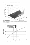
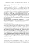
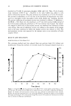
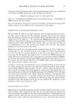
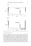
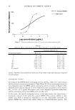
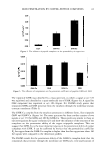
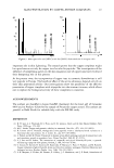
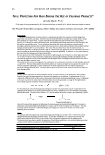
SKIN PENETRATION BY COPPER-PEPTIDE COMPLEXES 63 ml/ min -1, and the eluate was monitored at 2 00 nm using the UV /Vis detector. The concentration of peptide was determined by measuring the peak area. Electrospray MS was applied to identify copper complexes present in the acceptor cell. ESI-MS spectra were acquired in the range of 150-1500 µ using 20 ms dwell time and 0.1 µ of step size. The ion spray voltage of 4000 V was applied for positive and negative ion acquisition. The orifice potential was established at 80 V, as the one offering the best signal intensity and causing partial fragmentation of the molecular ion at the peptide bounds (34). DATA ANALYSIS To calculate the permeability coefficient, the cumulative amount of copper ions was plotted against the flux CT) of a compound across the membrane, determined at steady state (35). The permeability coefficient of the Cu2+ ion in the lipid membrane K P (cm·s-1) was calculated by Fick's first law of diffusion. Figure 1 shows exemplary permeation profiles of ligands and the amount of copper vs time. RESULTS AND DISCUSSION The aim of our research was to determine the influence of ligands (peptides) on the permeation process of copper ions. First, our studies confirmed the ability of copper ions to penetrate the model membrane without the determination of a compound form (copper ions or copper complexes). Second, we investigated the concentration of peptides that permeated the membrane. Finally, from the obtained data and ESI-MS results we were able to establish the form in which copper and peptides permeate. PERMEABILITY COEFFICIENT STUDY The results introduced in Figure 2 reveal a high influence of complexing agent (GHK or GSH) on the permeability coefficient of copper ions. In all cases, the permeation rates of copper ions were lower than those obtained for complexed copper. For this reason, it may be concluded that the complexing agents (GHK and GSH) accelerate the migration of copper ions through the model membranes. As shown in Figure 2, the influence of peptide complexes on the permeation of copper ions has different levels the influence of GHK on copper ion penetration was confirmed to be twice as strong as that of GSH. Research determined the permeation coefficient of peptides from the copper complexes. The concentration of G HK and GSH in the acceptor cell was determined by reversed phase liquid chromatography (RPLC) with UV-VIS detection. In Figure 3 the compari son of the permeation coefficients of GHK and GSH peptides from the copper complexes is presented. The figure proves that GHK and GHK-Cu have very similar values. What is more, on the basis of Figure 3 the conclusion that tripeptide complexation of copper does not change the K P value of GHK may be drawn. The GSH values were different: the K P for GSH was higher than that for the GSH copper complex. Similar properties of the penetration abilities of the GHK peptides confirmed the thesis that the structure and high affinity to the lipid structures of the membrane strongly influence the per meation process. The permeation coefficients of the peptides are significantly lower than those of copper ions.
Purchased for the exclusive use of nofirst nolast (unknown)
From: SCC Media Library & Resource Center (library.scconline.org)