2007 ANNUAL SCIENTIFIC MEETING 89 presenting cells' take up from outside the cell, predominantly bacterial antigens. Helper T cells respond by producing molecules called cytokines which are the hormones of the immune system and act locally and distantly to regulate the function of cells derived from both the innate and adaptive arm of the immune system. For example, some antigens direct helper T cells to produce groups of cytokines that are pro inflammatory and activate the function of macrophages, innate immune system cells, whereas other antigens may stimulate helper T cells to produce cytokines that facilitate the production of antibodies by B lymphocytes or accelerate an allergic reaction. Immunologic Disease Despite it complexity and many points of control, some individuals in the population develop diseases that are the result of immunologic 'hypersensitivity'. One example of immunologic hypersensitivity is the development of allergy which affects almost 20% of the population. Underlying allergic responses is the production of a class of antibody, IgE, to environmental antigens which are referred to as allergens. The IgE molecules bind to IgE Receptors on mast cells found in mucosa! tissues and are crosslinked when they bind the allergens. Crosslinking of the receptors on mast cells results in the degranulation and release of chemical mediators, such as histamine, that cause the allergic symptoms. A second type of immunologic hypersensitivity is autoimmune disease in which tolerance to self antigens is lost and either an antibody-mediated or cell-mediated response to host cells or proteins develops. Finally, immunodeficiency syndromes, either congenital or acquired, result in susceptibility to a variety of pathogens depending on which component of the immune system is defective. Conclusion Thus the immune system is a finely balanced system designed to protect the individual against invasion and subsequent disease production by a myriad of infectious agents. It utilizes an early defense system of cells and molecules, innate immunity, to eliminate infectious microbes together with an adaptive immune system that has a fine specificity for foreign substances and responds more effectively with each subsequent exposure to a microbe to provide long-lasting immunity.

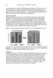
90 JOURNAL OF COSMETIC SCIENCE THE SKIN AS AN ENVIRONMENTALLY RESPONSIVE IMMUNOLOGICAL BARRIER John A. Wagner, Ph.D.1, Wanhong Ding2 and Richard D. Granstein2 1 Department of Neurology and Neuroscience, 2 Department of Dermatology, Weill Cornell Medical College, New York, NY 10021 Although the skin provides a vitally protective physical barrier, it provides an equally important immunological barrier to environmental pathogens. Antigen presenting cells that are resident in the skin can initiate and direct the organism's immune response. These antigen presenting cells are derived from the blood. They migrate into the skin, where they remain for extended periods in an immature state. Foreign antigens provide one of the signals that promote the maturation of these cells and their cells migration to lymph nodes. Despite the central role of exogenous antigens and adjuvants in this cascade, local signaling that occurs among cells within the skin play important regulatory roles that are just beginning to be elucidated. Here, we will focus on emerging evidence that signals derived from local nerves modulate the maturation and function of immune cells. We will also describe recent evidence that the nucleotide ATP acts as an endogenous adjuvant that signals the existence of tissue damage to the immune system. Cutaneous nerves are in intimate contact with Langerhans cells (one of the antigen presenting cells in the skin) and Langerhans cells express receptors for both a classic neurotransmitter (norepinephrine) and for at least three neuropeptides (CGRP, VIP, and PACAP). These neurotransmitters inhibit the ability of Langerhans cells to present antigen in vivo and in vitro. These effects occurred, at least in part, by modifying cytokine and chemokine production. At an intracellular level, many of the effects of neurotransmitters are mediated by an inhibition of the phosphorylation of IkappaB kinase, which prevents IkappaBalpha degradation and the subsequent activation of the transcription factor NF-kappaB. Thus, cutaneous immune responsiveness is modulated by neural activity While neurotransmitters are presumably derived from cutaneous nerves, ATP can be secreted by nerves, but ATP can also be released subsequent to tissue damage. Langerhans cells express several cell surface receptors for ATP. In contrast to neurotransmitter receptors, occupation of these receptors with either ATP or other purinergic agonists augments cutaneous immune responses, in part, by modulating the production of cytokines and co-stimulatory molecules. Activation of purinergic receptors results in activation of IkappaB kinase, increased IkappaBalpha degradation and activation NF-kappaB. Thus, the presence of ATP can be interpreted as a local indicator of tissue damage that is used to enhance immune responsiveness. To a first approximation, ATP acts in opposition to neurotransmitters in both a formal and a mechanistic sense, but there are many implications of this model that remain to be explored. It is clear that local signaling systems within the skin modulate immune responsiveness, and these signaling systems provide therapeutic targets that may be exploited to modulate cutaneous immunity
Purchased for the exclusive use of nofirst nolast (unknown)
From: SCC Media Library & Resource Center (library.scconline.org)