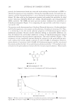
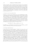
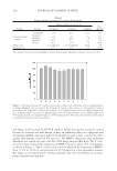
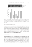
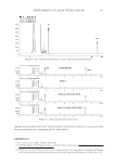
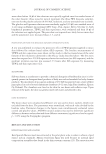
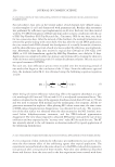
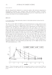
JOURNAL OF COSMETIC SCIENCE 256 acidifi cation of polar domains present within the hydrophilic lipid bilayers or by activation of acid protease crucial for desmosomal degradation (6). Furthermore, it has been reported that AHAs with a small molecular size are more active because they penetrate the skin more deeply (11). Glycolic acid is the simplest AHA, and it has the smallest molecular weight and size, followed by lactic, malic, tartaric, and citric acids, etc. This may explain the fact that glycolic acids tend to better penetrate the skin and accelerate skin regenera- tion. Mandelic acid and the organic acids contained in grape juice (tartaric acid, malic acid, citric acid, lactic acid, gluconic acid, and shikimic acid) have a greater molecular size and have more diffi culty in penetrating the skin. On the other hand, since the chemical peeling produces an insult to the skin, several side effects, such as erythema and redness, develop after treatment with exfoliating agents (3,9). Moreover, recent studies report that short-term dermal exposure with low concen- trations of exfoliating agents results in increased photosensitivity to UV light, measured as increased erythema and tanning (13,24). Skin tolerance to the exfoliating treatment is usually assessed by a visual and subjective record system. However, when objective and quantitative data are required, an instru- mental and non-invasive method is preferred for more accurate evaluations of the adverse skin effects. To this end, the skin tolerance and the photosensitizing effects of exfoliating acids were investigated by refl ectance spectrophotometric in vivo evaluation of skin ery- thema induced by topical application and after UV-light exposure. Since erythema is due to an increment in blood count in the subpapillary plexus of the skin and none of the main skin chromopheres (hemoglobin and melanin) absorb in narrow bands, the ery- thema index is not exclusively a linear function of hemoglobin content, but is affected by skin melanin content (25). On the basis of these assumptions, the skin refl ectance spectra, obtained by recording information on the optical spectrum of visible light ranging from 400 nm to 700 nm, is regarded as an accurate and reliable evaluation of the skin hemo- globin amount. Thereafter, the skin refl ectance spectral values permit calculation of the erythema index by subtracting the main melanin absorption peaks (510 and 610 nm) from the hemoglobin absorption peaks at wavelengths of 540 nm, 560 nm, and 580 nm Figure 4. Increase in skin sensitivity to UVB irradiation expressed by photosensitivity percentage after short-time treatment (four weeks) with GLY (glycolic acid), MAN (mandelic acid), and GA (grape acids) formulations containing 10% of acids vs control (no topical treatment). The photosensitivity % values were calculated from the erythema index obtained 24 hours after UVB exposure for each subject participating in the study.

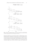
EFFICACY AND TOLERANCE OF EXFOLIATING AGENTS 257 (19). To better evaluate the skin tolerance of the tested acids, three different concentrations commonly used in light, medium, and deep peeling were topically applied. The study pointed out that skin erythema induced by topical application of the test organic acids in- creases with higher concentrations. Moreover, the spectrophotometric refl ectance approach is also able to evaluate slight changes in mild erythema induced by UV exposure. Recent experimental studies have demonstrated that subclinical irritation may be associ- ated with topical exposure to glycolic acid. It is conceivable that the pro-infl ammatory mediators released in irritated skin can affect events leading to UV-light sensitivity (13), increasing the risks of acute and chronic skin reactions. However, it is important to note that the rapid penetration of glycolic acid induces a more rapid skin exfoliation but causes a more intense skin redness and irritation. Thus, it is possible to infer that because of their molecular size, mandelic acid and grape acids are absorbed at a slower rate than glycolic acid, thereby causing less skin irritation. In addition, our results suggest that the spectro- photometric refl ectance approach used in this study represents a sensitive method for monitoring and comparing the effectiveness and the safety of different topical agents in skin exfoliating treatments. The methodologies and protocols used in this study may help in choosing the most appropriate topical agents for short/long term and mild/strong skin-exfoliating treatments. REFERENCES (1) W. E. Roberts, Chemical peeling in ethnic dark skin, Dermatol. Ther., 17, 196–205 (2004). (2) F. Furukawa and Y. Yamamoto, Recent advances in chemical peeling in Japan, Dermatol., 33, 655–661 (2006). (3) A. U. Bari, Z. Iqbal, and S. B. Rahman, Tolerance and safety of superfi cial chemical peeling with sali- cylic acid in various facial dermatoses, Indian J. Dermatol. Venereol. Leprol., 71, 87–90 (2005). (4) G. D. Monheit, Chemical peels, Skin Ther. Lett., 9(2), 6–11 (2004). (5) N. Zakopoulou and G. Kontochristopoulos, Superfi cial chemical peels, J. Cosmet. Dermatol., 5(3), 246– 253 (2006). (6) M. Fartasch, J. Teal, and G. K. Menon, Mode of action of glycolic acid on human stratum corneum: Ultrastructural and functional evaluation of the epidermal barrier, Arch. Dermatol. Res., 289, 404–409 (1997). (7) T. C. Flynn and W. P. Coleman, Topical revitalization of body skin, J. Eur. Acad. Dermatol. Venereol., 14, 280–284 (2000). (8) L. H. Couch and P. C. Howard, Quantifi cation of the glycolic acid in cosmetic products using reversed phase high performance liquid chromatography, Int. J. Cosmet. Sci., 24, 89–95 (2002). (9) C. Grover and B. S. Reddu, The therapeutic value of glycolic acid peels in dermatology, Indian J. Der- matol. Venereol. Leprol., 69, 148–150 (2003). (10) R. R. Gupta, B. B. Mahajan, and G. Garg, Chemical peeling—Evaluation of glycolic acid in varying concentrations and time intervals, Indian J. Dermatol. Venereol. Leprol., 67, 28–29 (2001). (11) S. M. Javaheri, S. Handa, I. Kaur, and B. Kumar, Safety and effi cacy of glycolic acid facial peel in Indian women with melasma, Int. J. Dermaytol., 40, 354–357 (2001). (12) N. Cassano, G. Alessandrini, M. Mastrolonardo, and G. A. Vena, Peeling agents: Toxicological and al- lergological aspects, J. Eur. Acad. Dermatol. Venereol., 13(1), 14–23 (1999). (13) K. Kaidbey, B. Sutherland, P. Bennett, W. G. Wamer, C. Barton, D. Dennis, and A. Kornhauser, Topi- cal glycolic acid enhances photodamage by ultraviolet light, Photodermatol. Photoimmunol. Photomed., 19, 21–27 (2003). (14) C. Ostacolo, A. Sacchi, A. Bernardi, S. Lanieri, A. Brunetta, and G. Pantini, Perfl uoropolyether phos- phate: Skin exfoliation after a topical pre-treatment, TEWL and skin elasticity, by in-vivo non-invasive methods, Int. J. Cosmet. Sci., 29, 391–398 (2007). (15) G. Pantini, R. Ingoglia, F. Brunetta, and A. Brunetta, Sunless tanning products containing dihydroxy- acetone in combination with a perfl uoropolyether phosphate, Int. J. Cosmet. Sci., 29, 201–209 (2007).
Purchased for the exclusive use of nofirst nolast (unknown)
From: SCC Media Library & Resource Center (library.scconline.org)