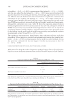
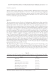
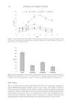
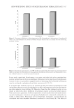
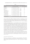
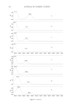
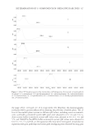
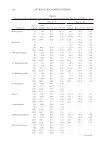
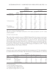
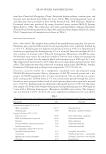
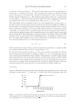
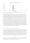
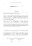
JOURNAL OF COSMETIC SCIENCE 300 minute (cpm), in cultures treated with 1.25 μM AE 7647 ± 2 423 cpm, in cultures treated with 2.5 μM AE 4680 ± 1535 cpm, and in those treated with 5 μM AE 1815 ± 566 cpm (mean ± SEM). The results of ANOVA showed that AE significantly inhibited keratinocyte proliferation (Figure 1A) in a dose dependent manner ( p = 0.019), with the maximal inhibitory effect achieved at 5 μM AE ( p = 0.021 compared to control). Al- though AE was able to signifi cantly inhibit keratinocyte proliferation at 5 μM concen- tration, considerable differences in keratinocyte response were observed among the cultures obtained from different individuals (Figure 1A). The observed individual differences were confi rmed by comparing the IC50 values between different donors (Table I), which revealed that keratinocytes from some donors display markedly differ- ent sensitivity to the antiproliferative action of AE (e.g., compare donors No. 8 and 10). The observed antiproliferative effect of AE was not accompanied by a signifi cant in- crease in LDH release during a 72 h incubation period (Figure 1B) (ANOVA p = 0.66), as well as at earlier time points (6, 24, and 48 h data not shown), indicating that the inhibitory effect of the drug on keratinocyte proliferation was probably not due to ne- crotic cell death. THE INFLUENCE OF AE ON KERATINOCYTE APOPTOSIS In order to explore the mechanisms underlying the observed inhibition of keratinocyte proliferation, we assessed whether AE induces apoptotic cell death (Figure 2). A signifi - cant increase in the percentage of apoptotic (TB−/FDA−) keratinocytes could be observed 6 h upon addition of 5 μM AE (16.7 ± 1.3, mean ± SEM) compared to the untreated cultures (10.4 ± 0.1, mean ± SEM, p = 0.001) (Figure 2). The number of necrotic cells in keratinocyte cultures did not signifi cantly change upon treatment with AE (Figure 2), thus further supporting results of the LDH release assay (data not shown). DISCUSSION In this study, we demonstrated antiproliferative effect of AE on the primary human kera- tinocytes from 10 different donors cultivated in vitro (Figure 1A). The antiproliferative effect of AE was previously reported in various human cell cultures at 20–80 μM concen- tration (6). It was a consequence of induction of p21, p53 and subsequent G1 cell cycle arrest (7), or increased number of cells within S phase (8). In some experiments, increased proportion of cells cycling at a higher ploidy level ( G2/M) was observed (8). The latest may explain increased DNA synthesis in the rat hepatocytes reported by Wolfl e et al. in this particular experimental setup (4). In line with previously presented data (6), our Table I The Half Maximal Inhibitory Conc entrations of Aloe-emodin for Proliferation of Keratinocyte Cultures from 10 Different Donors Sample 1 2 3 4 5 6 7 8 9 10 Mean SEM IC50 (μM) 2.96 3.91 3.02 3.46 2.86 2.14 1.79 0.96 3.22 5.40 2.97 0.38 IC50 ( ppm) 1.10 1.45 1.12 1.28 1.06 0.79 0.66 0.36 1.19 2.00 1.10 0.14

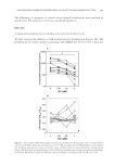
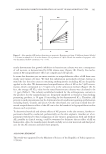
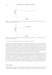
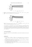
301 ALOE-EMODIN INHIBITS PROLIFERATION OF ADULT HUMAN KERATINOCYTES results demonstrate that growth inhibition in keratinocyte cultures was not a consequence of cell necrosis, as demonstrated by LDH release assay (Figure 1B). Clearly, the mecha- nisms of AE antiproliferative effect are complex and cell-specifi c. It seems that keratinocytes are more sensitive to antiproliferative effect of AE than com- monly used tumor cell lines. We fi nd this information particularly relevant, having in mind the fact that aloe extracts containing anthraquinones including AE are frequently applied to the skin. We demonstrated antiproliferative effect of AE at the 5 μM concen- tration, which corresponds to 1.35 ppm w/w in the cultivation medium (Figure 1A). In fact, the average AE IC50 value for the tested keratinocyte cultures was calculated to be 1.1 ppm (Table I). The industry-established standard for the anthraquinone contents in the products for the nonmedicinal use, frequently marketed as wound or burn healing promoting products, is 50 ppm (3) exceeding by far demonstrated IC50. Therefore, AE present in such preparations may be detrimental in all conditions requiring epithelization including burns, wounds, and ulcers. On the other hand, one can keep in mind that ob- served antiproliferative effect of the AE can in fact be benefi cial in hyperproliferative skin diseases such as psoriasis. To determine benefi cial and adverse effects of AE present in the aloe extracts, further ex- periments should be conducted, preferentially on the skin explants. Another set of ex- periments with head-to-head comparison of aloe extracts preparations with and without AE, possibly in clinical setting, could be warranted to delineate the net effect of AE on human skin. Also, the manufacturers should carefully screen raw materials and fi nal prod- ucts for the AE contents and label them properly. ACKNOWLEDGMENT This work was supported by the Ministry of Science of the Republic of Serbia (grant no. 175038). Figure 2. Aloe-emodin (AE) induces keratinocyte apoptosis. Keratinocytes from 10 different donors (labeled 1–10) were incubated for 6 h in the absence (0) or presence of 5 μM AE and the number of apoptotic cells was determined by fl ow cytometry. ∗∗p 0.01.
Purchased for the exclusive use of nofirst nolast (unknown)
From: SCC Media Library & Resource Center (library.scconline.org)