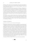
RELEASE OF C. ALBICANS FROM SKIN 91 contribute substantially to the attachment of soil or yeast to the skin. Electrostatic inter- actions occur between charge density and distribution on a soil’s surface and inversely charged components on another surface (17,19). Examples of electrostatic interactions include dipole–dipole forces, hydrogen bonds, cationic, and anionic interactions (4,16,22). Molecules with dipole moments attract each other electrostatically by aligning their pos- itive and negative ends in close proximity (23). Hydrogen bonding occurs when a hydro- gen atom is covalently bonded to an electronegative atom and also attracts an additional electronegative atom (5). The attraction created by individual dipole moments and hy- drogen bonding should produce only small changes however, the cumulative effects of simultaneous events may generate signifi cant changes. The biochemistry and structure of biological membranes cause them to be negatively charged (18) therefore, a cationic exchanger should repel yeast and an anionic exchanger should attract yeast, promoting removal from skin. Charge interactions are important to skin cleansing because alteration of the charge affi nity between the soil and cleaning web or solution can increase cleaning effectiveness. This paper describes a method for use of the cationic exchanger carboxymethylcellulose (CMC) to promote enhanced removal of C. albicans from human skin. MATERIALS AND METHODS YEAST CULTURE C. albicans (ATCC 10231) was subcultured using Sabouraud Dextrose (SAB-DEX) me- dium (Becton Dickinson, Cockeysville, MD) overnight at 37°C. The following day, 20 ml SAB-DEX broth was inoculated with 2–3 isolated C. albicans colonies and incu- bated at 32°C for 18 h with shaking at 220 rpm. The broth culture was diluted to 1 × 10⁵ colony-forming units (CFU)/ml with 50 mM sterile potassium phosphate buffer pH 7.2 (VWR Industries, Batavia, IL). VISUAL RELEASE PROTOCOL The following protocol, outlined in Figure 1, was used to measure the effectiveness of various test materials (described in Table I) to induce release of C. albicans adhered to skin tape. Skin tape strips were produced by pulling D-Squame skin-sampling discs (CuDerm Corporation, Dallas, TX) four times from adjacent adult male volar forearm sites. The skin tape strips were placed into deep six-well plates (Becton Dickinson, Franklin Lakes, NJ), and the exposed adhesive was blocked with 2.0 ml of 5% bovine serum albumin (BSA Sigma, St. Louis, MO) in phosphate buffered saline (PBS 150 mM NaCl, 50 mM potassium phosphate, pH 7.4) for 60 min at 33°C while shaking at 220 rpm. The fl uid was removed from each well, the wells washed three times with 50 mM potas- sium phosphate, and 1.0 ml (105 CFU/ml) of C. albicans solution, prepared as described in the previous section, was added to each tape strip. Next, 1.0 ml of trypticase soy broth (TSB Difco Labs, Detroit, MI) was added to each tape strip and the plates were incubated at 33°C for 60 min. The fl uid was aspirated and the tape strips were washed three times with 3.0 ml tris(hydroxymethyl)aminomethane (TRIS)-buffered saline (TBS 50 mM

JOURNAL OF COSMETIC SCIENCE 92 Tris base, 150 mM NaCl, pH 7.4). Both sides of each tape strip were rinsed with a stream of TBS and placed in fresh 6-well plates. Test materials were suspended in TBS and 2.0 ml was added to the designated tape strip. The tape strips were incubated for 15, 30, or 60 min at 25°C while shaking at 220 rpm. The tape strips were then rinsed three times with 3.0 ml deionized (DI) water. Each tape strip was fi xed by soaking in 2.0 ml of 2.5% glutaraldehyde (Sigma) for 10 min. The tape strips were then rinsed three times Table I Materials Used in Experiments Material Use concentration (mg/ml) Exchange capacity Size Supplier Benonite 8.0 NAa Powder Sigma, St. Louis, MO Carboxymethyl (CM)-Cellulose 8.0 0.6 meq/g Fibrous Sigma, St. Louis, MO Cellulose 8.0 NA Fibrous Sigma, St. Louis, MO Diethylaminoethyl (DEAE)-Cellulose 8.0 1 meq/g Powder Sigma, St. Louis, MO Diethyl- (2-hydroxypropyl) aminoethyl (QAE–Cellulose 8.0 0.92 meq/g Fibrous Sigma, St. Louis, MO Cellulose-Phosphate 8.0 3.8 meq/g Powder Sigma, St. Louis, MO Chitin 8.0 NA Powder Sigma, St. Louis, MO Carboxymethyl (CM)-Cellulose 0.4, 0.25 50–70 mg Lysozyme/g 3–4 μm SCIGEN, Kent, UK Polyethylenimine (PEI)-Cellulose 0.4 70–100 mg Albumin/g 3–4 μm SCIGEN, Kent, UK Diethylaminoethyl (DEAE)-Cellulose 0.4 70–100 mg Albumin/g 3–4 μm SCIGEN, Kent, UK a NA = Not Applicable. Figure 1. Diagram of yeast attachment assay.
Purchased for the exclusive use of nofirst nolast (unknown)
From: SCC Media Library & Resource Center (library.scconline.org)