FUCUS EXTRACT: COSMETIC TREATMENT FOR UNDER-EYE DARK CIRCLES 107 WESTERN BLOT Cells were grown to confl uence and then treated with tested product in their respective medium for 3 days for NHEK cell and for 24 h for NHDF cells at 37°C with 5% CO2. After rinsing once with PBS, cells were processed for Western blotting by lysis in RIPA buffer containing a cocktail of protease inhibitors. The lysate was sonicated for 15 s, cen- trifuged 10 min at 12000g, and the supernatants were quantifi ed for protein content by BCA assay. HO-1 protein levels were then analyzed by electrophoresing 30 μg cellular protein in a 4%–20% acrylamide gradient gel (Bio-Rad, Hercules, CA), transferring to a nitrocellulose membrane and blotting 90 min with mouse anti-human HO-1 antibodies and mouse anti-β-actin antibodies (to normalize protein loading). After washing, the membrane was blotted with HRP-linked anti-mouse antibodies for 60 min. HO-1 and β-actin bands were resolved and quantifi ed by chemiluminescence on a Kodak Image Sta- tion 4000R. Cell treatments, extractions, protein quantifi cations, and Western blots were performed in triplicate. DPPH ASSAY Fucus vesiculosus water extract was diluted to 0.5%, 1%, 2%, and 5% in PBS. Trolox was dissolved in ethanol and diluted to concentrations of 2.5, 5, 10, 20, 30, 40, and 50 μM, which is used to generate a standard curve. DPPH was dissolved in ethanol to make 100 μM solution. In a 96-well plate, 100 μL of sample or standard solutions was added to wells in six replicates, and then 100 μL of DPPH solution was added to triplicate wells, and 100 μL of ethanol without DPPH to the other triplicate wells for background reading. After mixing at room temperature for 20 min, absorbance was read at 510 nm. In order to minimize background interference for colored samples, the background reading is sub- tracted from the fi nal absorbance of these samples. IL-8 ASSAY Cells were pretreated for 4 h with basal medium (Invitrogen, medium 154 with no growth supplement). Then, cells were incubated overnight in treatment media (Invitro- gen, medium 154 with 10 ng/ml of IL-β and tested product added) at 37°C with 5% CO2. The level of IL-8 released into culture medium was measured by ELISA method. In brief, mouse anti-human IL-8 was coated overnight in a 96-well plate. Samples and stan- dard were then added to the plate and incubated for 2 h. After washing, a biotinylated goat anti-human IL-8 was added to the plate and incubated for 2 h. A streptavidin- conjugated horseradish peroxidase and substrate was used to measure the amount of IL-8 content by recording the optical density at 450 nm with correction at 540 nm. COLLAGEN I ASSAY—DELFIA METHOD Collagen I was assayed using a method developed internally (Immunoassay method for in vitro measurements, patent application fi led). After treatment with the products, cells

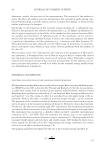
JOURNAL OF COSMETIC SCIENCE 108 were disrupted with dedicated lysis solution. This solution allows the disruption of cell membranes without solubilizing the deposited matrix. The deposited collagen I was de- tected by primary antibody anti-collagen I (Interchim, Montlucon, France) and with the secondary antibody anti-IgG coupled to a DELFIA® Europium (Perkin Elmer, Courta- boeuf, France) probe. Europium-related fl uorescence (λexc, 340 and λem, 615 nm), propor- tional to the quantity of deposited collagen was measured. STATISTICAL ANALYSIS The statistical analysis (two-tailed) for in vitro studies were performed using Student’s t-test and analysis of variance on the multiple comparisons with a threshold of signifi - cance set to 5% ( p 0.05). RESULTS AND DISCUSSION EFFECT OF FUCUS EXTRACT ON HO-1 STIMULATION IN VITRO Skin responds to environmental assault and endogenous factors by changing the vascular integrity, leading to the blood leakage and heme accumulation in the surrounding tissue space and causing dark circles. In this study, experiments were performed to evaluate the effect of Fucus extract in the HO-1 stimulation activity in cultured cells. The HO-1 stimulation effi cacy of Fucus extract is evidenced fi rst at the gene level by RT- PCR (Figure 3). For initial screening, both fi broblasts and keratinocytes were treated with 5% of the Fucus extract in their respective medium for 24 h at 37°C with 5% CO2. Total RNA was isolated, cDNA was synthesized and used as template, and a probe set specifi c to HO-1 gene was used for this reaction. The HO-1 mRNA expression profi le was compared to non-treated control, and the results from both cell types are described in Table I. Figure 3. Fucus extract stimulates heme oxygenase-1 gene expression in keratinocytes- (Log base is 2, 1log = 2 fold) Mean ± SD on 3 assays – Paired Student’s t- test - **: p 0.01- ***: p 0.001 compared to control.
Purchased for the exclusive use of nofirst nolast (unknown)
From: SCC Media Library & Resource Center (library.scconline.org)