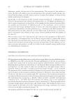
JOURNAL OF COSMETIC SCIENCE 92 Tris base, 150 mM NaCl, pH 7.4). Both sides of each tape strip were rinsed with a stream of TBS and placed in fresh 6-well plates. Test materials were suspended in TBS and 2.0 ml was added to the designated tape strip. The tape strips were incubated for 15, 30, or 60 min at 25°C while shaking at 220 rpm. The tape strips were then rinsed three times with 3.0 ml deionized (DI) water. Each tape strip was fi xed by soaking in 2.0 ml of 2.5% glutaraldehyde (Sigma) for 10 min. The tape strips were then rinsed three times Table I Materials Used in Experiments Material Use concentration (mg/ml) Exchange capacity Size Supplier Benonite 8.0 NAa Powder Sigma, St. Louis, MO Carboxymethyl (CM)-Cellulose 8.0 0.6 meq/g Fibrous Sigma, St. Louis, MO Cellulose 8.0 NA Fibrous Sigma, St. Louis, MO Diethylaminoethyl (DEAE)-Cellulose 8.0 1 meq/g Powder Sigma, St. Louis, MO Diethyl- (2-hydroxypropyl) aminoethyl (QAE–Cellulose 8.0 0.92 meq/g Fibrous Sigma, St. Louis, MO Cellulose-Phosphate 8.0 3.8 meq/g Powder Sigma, St. Louis, MO Chitin 8.0 NA Powder Sigma, St. Louis, MO Carboxymethyl (CM)-Cellulose 0.4, 0.25 50–70 mg Lysozyme/g 3–4 μm SCIGEN, Kent, UK Polyethylenimine (PEI)-Cellulose 0.4 70–100 mg Albumin/g 3–4 μm SCIGEN, Kent, UK Diethylaminoethyl (DEAE)-Cellulose 0.4 70–100 mg Albumin/g 3–4 μm SCIGEN, Kent, UK a NA = Not Applicable. Figure 1. Diagram of yeast attachment assay.

RELEASE OF C. ALBICANS FROM SKIN 93 with 3.0 ml of DI water, and stained with 0.5 ml calcofl uor white (Difco, Ann Arbor, MI) for 10 to 15 min. The tape strips were again rinsed three times with 3.0 ml DI water and allowed to air dry. Once the tape strips were air-dried, the yeast cells were enumerated visually with an Olympus BH2 fl uorescent microscope (Olympus Corporation, Lake Success, NY) fi tted with a 405 nm excitation fi lter and a 455 nm barrier fi lter. The tape strips were placed with the white crescent label near the bottom edge of a microscope slide, perpendicular to the microscope objective. A 20X objective was used and the slide was positioned so that the fi eld of view bisected the tape strip. Only the cells appearing within the fi eld of view (an area of approximately 2 × 107 μm2) were counted. The fi eld of view represented 5% of the total tape strip area. The percent removal was calculated as follows: number of yeast on control tape strip number of yeast on treated tape strip number of yeast on control tape strip q100 This method estimated approximately 104 yeast cells bound to a 22-mm diameter D-Squame tape strip under the described conditions. Tape strips were evaluated in trip- licate for each condition. Digital photomicrographs were taken with a SPOT Digital Camera (Diagnostic Instruments, Sterling Heights, MI). VIABLE COUNT RELEASE PROTOCOL Skin tape strips produced using the previously described procedure were used to assess the ability of 3–4 μm CMC particles (described in Table I) to release C. albicans from the skin strips. The exposed adhesive on the tape strips was blocked with 2.0 ml of 5% bovine serum albumin (BSA) in PBS and incubated for 60 min at 33°C while shaking at 220 rpm. After the blocking solution was removed, each well was washed three times with potas- sium phosphate buffer (KP), and then 1.0 ml of C. albicans suspension (105 CFU/ml) and 1.0 ml of TSB were added to each tape strip. The plates were incubated at 33°C for 60 min, the fl uid was aspirated, and the tape strips were rinsed three times with 3.0 ml TBS. A suspension of 3–4 μm CMC (2.0 ml 0.25 mg/ml in TBS) was added to each tape strip and 2.0 ml TBS was added to the control. The tape strips were incubated at 25°C while shaking at 220 rpm. At 0, 15, 30, and 60 min after addition of the CMC suspension, fl uid samples were collected from each well (in triplicate) and plated on SAB-DEX agar. The agar plates were then incubated for 24 h at 33°C and the resulting yeast colonies were counted to determine the numbers viable of yeast in each solution. EFFECT OF PH ON RELEASE EFFICACY To determine the potential effect of pH on the release C. albicans from the skin by bath tissue containing CMC particles, skin tape strips were prepared as described previously. The exposed adhesive on the tape strips was then blocked with 2.0 ml of 5% BSA in PBS for 60 min
Purchased for the exclusive use of nofirst nolast (unknown)
From: SCC Media Library & Resource Center (library.scconline.org)