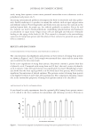
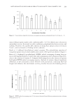
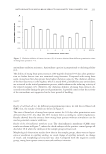
J. Cosmet. Sci., 65, 217–224 (July/August 2014) 217 Internal structure changes of eyelash induced by eye makeup KEN-ICHI FUKAMI, TAKAFUMI INOUE, TOMOMITSU KAWAI, MUNEKI SAKATA, MASAHIRO NAGANO, KOUJI TAKEHARA, AKIHISA TAKEUCHI, KENTARO UESUGI, and YOSHIO SUZUKI, Skincare Research Laboratory (K.F., T.K., K.T.), Innovative Beauty Science Laboratory (T.I.), Make-up Research Laboratory (M.S., M.N.), Kanebo Cosmetics Inc., Kotobuki-cho 5-3-28, 250-0002, Japan, and Japan Synchrotron Radiation Research Institute, Koto 1-1-1, Hyogo 679-5198 (A.T., K.U., Y.S.), Japan. Accepted for publication May 27, 2014. Synopsis To investigate how eye makeup affects eyelash structure, internal structure of eyelashes were observed with a scanning X-ray microscopic tomography system using synchrotron radiation light source. Eyelash samples were obtained from 36 Japanese women aged 20–70 years and whose use of eye makeup differed. Recon- structed cross-sectional images showed that the structure of the eyelash closely resembled that of scalp hair. The eyelash structure is changed by use of eye makeup. There was a positive correlation between the fre- quency of mascara use and the degree of cracking in cuticle. The positive correlation was also found between the frequency of mascara use and the porosity of the cortex. By contrast, the use of eyelash curler did not affect the eyelash structure with statistical signifi cance. INTRODUCTION Whether eye makeup induces characteristic changes in the structure of eyelashes is of interest. The growth cycle of the eyelash is approximately 90 days (1), which is shorter than that of scalp hair of two to six years (2). Therefore, there would appear to be less opportunity for eyelashes to be damaged than scalp hair. It has been re- ported that characteristics of eyelashes are not infl uenced by eye makeup use (3). However, the makeup treatments that are applied to the eyelash differ from those that are applied to the scalp hairs, e.g., mascara, eyelash curler, false eyelashes, and eyelash extensions. Eyelashes might be damaged by these treatments. In this study, we investigated the effects of mascara and eyelash curler use on eyelash internal structure. Address all correspondence to Takafumi Inoue at inoue.takafumi@kanebocos.co.jp.

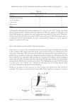
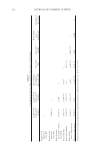
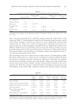
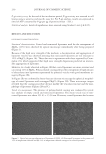
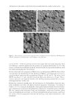
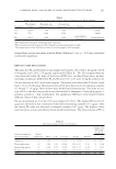
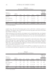
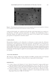
JOURNAL OF COSMETIC SCIENCE 218 The internal structures of the eyelashes were observed using a differential phase-contrast scanning X-ray microscopic computed tomography (CT) system (4). This system enables to observe density distribution of intact samples by measuring the phase gradient of transmitted X-ray probe. MATERIALS AND METHODS EYELASH SAMPLES Eyelash samples were obtained from 36 Japanese women whose use of eye makeup dif- fered. A questionnaire was used to determine their age and whether the women used mascara and/or an eyelash curler, and if so, with what frequency in last two months. The number of women in each age category is shown in Table I, and the frequencies of two eye makeups are shown in Table II. OBSERVATION OF EYELASH STRUCTURE The measurement was performed at the undulator beamline 20XU of SPring-8 (Hyougo, Japan). We employed a differential phase-contrast scanning X-ray microscopic CT system (4), which has been developed to observe the internal structure of human hair sample (5). The differential phase-contrast image representing the variation of refractive index of sample is obtained by measuring the angle of defl ection of the transmitted X-ray beam. The decrement of refractive index from unity is approximately proportional to the elec- tron density. The electron density is generally proportional to mass density. The samples were set in the air. So, the phase-contrast image reconstruction by the CT system corre- sponds to the mass density map of eyelash samples. The sample setup of the scanning X-ray microscopy system is shown in Figure 1. The eyelash sample was positioned such that the eyelash bulb was uppermost, and regions of the eyelash shaft located 2 mm from the eyelash bulb were scanned. The eyelash samples were observed in the atmosphere. More details of the experimental setup are described elsewhere (4). A focused 8 keV X-ray beam with a diameter of 100 nm was used as a scan probe. The transversal scan pitch was 100 nm. For a CT measurement, transversal scan was repeated Table I Age Distribution of Volunteers Age (years) Number of volunteers 20–30 7 30–40 8 40–50 8 50–60 8 60–70 5
Purchased for the exclusive use of nofirst nolast (unknown)
From: SCC Media Library & Resource Center (library.scconline.org)