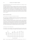
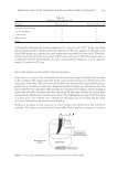
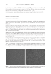
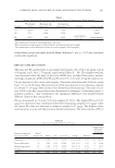
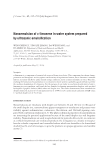
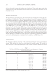
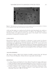
NEW RESISTANT LIPOSOME COATED WITH POLYSACCHARIDE FILM FOR COSMETIC APPLICATION 231 size of 514.00 ± 79.00 nm and thus are two times larger than noncoated liposomes. This size difference could be explained by the presence of a polysaccharide fi lm (11–16). The stearic acid from Stearoyl Inulin would be anchored inside the liposomal membrane with the inulin surrounding the outer bilayer. On the other hand, the entrapment of MgCl2 into coated liposomes (MCCL) affected their size and their size distribution. In the presence of MgCl2, coated liposome size was in- creased without impacting their morphology (Figures 2A, B, and C). The mean size of MCCL was about 1.37 μm, and 90% of vesicles had a size inferior to 3.14 μm. The in- crease in liposome size might be linked to the swelling of polysaccharide fi lm in contact with high amounts of salts (22). Beyond size parameter modifi cation, the introduction of a hydrophobic chain into lipo- some membrane may affect the physicochemical properties of liposomes. According to Carafa et al. (11), coating by hydrophobic chain grafted polysaccharides decreased transi- tion temperature of liposome phospholipids compared to nongrafted polysaccharides. We also supposed that hydrogen bonds and hydrophobic interactions between carbon chain of phospholipids and stearic acid from hydrophobized polysaccharides could limit the release of the encapsulated molecules by forming a very tight embedment in the membrane. Figure 2. Freeze-fracture electron microscopy (×41,100): (A) Empty noncoated liposomes, (B) Magnesium chloride entrapped in coated liposomes, and (C) Empty coated liposome.

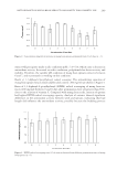
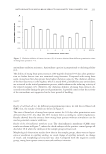
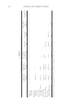
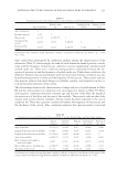
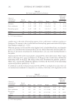
JOURNAL OF COSMETIC SCIENCE 232 STABILITY OF LIPOSOMES Resistance of coated and noncoated liposomes to surfactants. The physicochemical stability of empty coated and noncoated liposomes was evaluated in the presence of ionic (SDS, SLES) and nonionic surfactants (Triton X-100, Polysorbate 20, polyglyceryl-10-laurate, Behe- nyl alcohol ethoxylate). The maximum percentages of ionic surfactants which do not destabilize empty coated and noncoated liposomes are shown in Table I. The shape of liposomes was observed after each period of time (Days 1, 7, 15, and 30) by optical microscope (×1000) at different ionic surfactant concentrations (1%–10%). Coated liposomes resisted up to 3% of both ionic surfactants until 30 days of storage in the presence of SDS and SLES. How- ever, noncoated liposomes are less resistant and remained stable at 1% and 2% of SDS and SLES during the period of storage, respectively. These observations allowed us to conclude that the coating process increases the stability of liposomes against ionic surfactants. The effect of different nonionic surfactant concentration (1%–10%) on the stability and shape of empty coated and noncoated liposomes was summarized in Table II. Firstly, coated and noncoated liposomes are more resistant to nonionic surfactants than to ionic ones. Whatever the kind of nonionic surfactant, the coating process improved the stability of liposomes. Our results showed that coated liposomes remained stable with good integrity in the presence of 9% of polysorbate 20 and 9% of polyglyceryl- 10-laurate for 30 days. Coated liposomes resisted up to 9% of behenyl alcohol 25 EO for 15 days but only up to 6% after 30 days. However, Triton X-100 is the most solu- bilizable nonionic surfactant as coated liposomes were stable until 30 days of storage in the presence of 3% of Triton X-100. Noncoated liposomes are more resistant to behenyl alcohol 25 EO and polyglyceryl-10-laurate than to polysorbate 20 and Triton X-100. Surfactants are widely used for the solubilization of phospholipid membrane. Mady et al. (23) have reported that the solubilization process is divided into three stages. (i) Stage I: corresponds to the insertion of surfactants into the bilayer membrane. (ii) Stage II: reached when the phospholipid membrane is saturated with surfactants, and followed by the formation of micelles. (iii) Stage III: Mixed lipid-surfactant micelles enriched in surfactant. Table I Resistance Kinetics of Coated and Noncoated Liposomes to Ionic Surfactants during 30 Days of Storage at 25°C T0 Day 1 Day 7 Day 15 Day 30 Percentage of SDS Noncoated liposome 1.0 1.0 1.0 1.0 1.0 Coated liposome 4.0 4.0 4.0 4.0 3.0 Percentage of SLES Noncoated liposome 2.0 2.0 2.0 2.0 2.0 Coated liposome 4.0 4.0 4.0 3.0 3.0 SDS: Sodium dodecyl sulfate, SLES: Sodium lauryl ether sulfate.
Purchased for the exclusive use of nofirst nolast (unknown)
From: SCC Media Library & Resource Center (library.scconline.org)