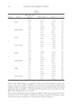
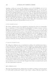
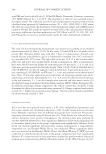
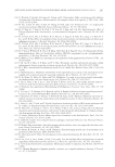
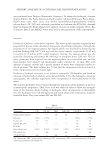
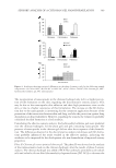
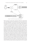
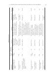
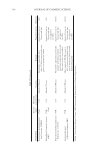
JOURNAL OF COSMETIC SCIENCE 282 challenged cellular environment, the fi nding that GSSG is the same in both cell types raises the possibility that eumelanin-producing melanocytes may normally utilize less GSH for their cellular functions. Ferritin. As iron plays a signifi cant role in the generation of ROS, we next measured the levels of ferritin, a storage protein of bioavailable iron, in both types of cells. Because serum is an exogenous source of iron in cell culture, we were careful to standardize both cell lines in 1% serum. As shown in Figure 4, ferritin levels in melanocytes were nearly fourfold higher than in NHEKs. Thus, NHEKs contained 23.3 ng (±5 SEM) ferritin/mg protein, whereas melanocytes contained 97.1 ng (±6 SEM) ferritin/mg protein. To further understand this difference in the context of other cells, we compared ferritin levels in other cell types. MCF-10a cells, immortalized mammary epithelial cells, had 13.0 ng Figure 3. GSH and GSSG were measured in NHEKs and melanocytes using a chemiluminescence-based assay. GSH levels were lower in melanocytes than in NHEKs but GSSG levels were both 12.2% of the total GSH. A linear regression curve was also developed with GSH standards to determine cell molar concentra- tions and then normalized to cell number as reported in the text. Data are expressed as mean ± SEM. Figure 4. Ferritin concentrations were assayed using an ELISA technique and showed increased levels of ferritin in melanocytes in comparison to NHEKs, MCF-10, as well as in HepG2, an iron-rich hepatocarci- noma cell. Data are expressed as mean ± SEM.

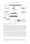
ENDOGENOUS DNA OXIDATION CORRELATES TO INCREASED IRON LEVELS IN MELANOCYTES 283 (±3 SEM) ferritin/mg protein. Intriguingly, even HepG2 cells, hepatocarcinoma cells derived from iron-enriched liver tissue, were signifi cantly lower than melanocytes at 33.2 ng (±2 SEM) ferritin/mg protein. These results demonstrate that melanocytes appear to have higher levels of iron in comparison to other cell types and support the concept of a higher oxidative environment in melanocytes. DISCUSSION Previously, we reported a fourfold higher level of H2O2 in NHEKs than in melanocytes (6), which should indicate less oxidative stress in melanocytes. We further showed that H2O2 could permeate by passive diffusion from NHEKs to melanocytes. Nevertheless, despite these previous fi ndings, we now report higher levels of oxidative DNA in mela- nocytes as compared to NHEKs, as well as lower concentrations of GSH, both of which are well-established biomarkers of oxidative status. Because NHEKs and melanocytes had similar UVB-induced 8-oxo-dG potential and because higher levels of 8-oxo-dG were also found in mouse-derived melanocytes, our data suggest that elevated states of oxidation may be characteristic for melanocytes. To account for these differences, we pro- pose that the higher levels of iron that we measured in melanocytes in this study contrib- ute to higher levels of 8-oxo-dG. A possible reaction sequence might involve a Fenton reaction between iron and H2O2 leading to the production of hydroxyl radicals and oxida- tive DNA. Because H2O2 is able to penetrate through membranes and because ferritin has been found in the nucleus (18), the possibility that H2O2 will react with ferrous ions inside the nucleus is suggested as a possible mechanism for increased melanocytic oxida- tive DNA. This reaction sequence might also explain, at least in part, the reduced amount of hydrogen peroxide that we previously determined in melanocytes. In this report, we show that melanocytes are signifi cantly higher in iron, compared not only to NHEKs but also to iron-rich hepatocarcinoma cells. As melanocytes are specialized epidermal cells dedicated to the synthesis and secretion of melanin in order to protect skin against UV-induced photo damage, the increased presence of iron raises the question as to why a melanocyte would tolerate such a high risk/benefi t iron ratio. Although ferritin seques- ters approximately 4500 ferric ions per molecule, it also releases iron as reactive ferrous ions (19). Iron ions have been shown to induce DNA base modifi cations in cellular DNA (20), and understanding this reaction dynamic in melanocytes may be an important factor in under- standing the melanogenic process. Intriguingly, 8-oxoguanine has also recently been de- scribed as a signaling molecule (21), and perhaps future research will link it to melanocyte gene activation, as well. Further, recent work by Gruber and Holtz (22) demonstrated increased levels of ferritin gene expression after treatment with conventional skin lighteners and their data also support the concept of iron fl ux in relation to melanocyte function. As postmenopausal women appear to have higher levels of cutaneous iron (11), our data suggest a possible correlation between this group and the development of age-related melanopathologies, such as lentigines. If future clinical studies support this hypothesis, then research should be targeted toward producing iron-reduction treatments (23). More- over, increased levels of melanocytic oxidative DNA due to increased iron may be a com- mon denominator that may help to explain the occurrence of melanomagenesis even in sun-protected areas of skin. In conclusion, higher levels of oxidative DNA appear to be present in normal human epidermal melanocytes and correspond to increased amounts of reactive bioavailable iron.
Purchased for the exclusive use of nofirst nolast (unknown)
From: SCC Media Library & Resource Center (library.scconline.org)