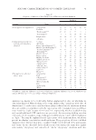
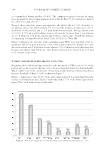
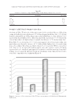
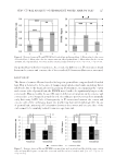
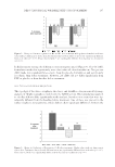
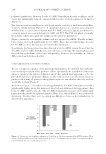
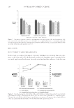
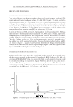
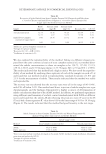
STRUCTURAL ANALYSIS OF PERMANENT WAVED HAIR BY SAXS 129 case of inclined IFs as seen in the orthocortex (12), swelling of IFAP longitudinally aligned the slope of the IFs, which led to the IF orientation becoming isotropic. Last, in the comparison of stretched SLS-immersed hair and permed hair, there were no differences in distance between IFs, whereas FWHM in the convex side of the curl in stretched SLS-immersed hair was signifi cantly smaller than that of the permed hair. Responsiveness on the convex side of the curl was more noticeable because of the difference in the stretching strain between the convex and concave sides of the curl when making the curl shapes (16). Therefore, it was conjectured that the change in the convex side was more noticeable when the strain was relieved. According to previous research, it was reported that IF orientation in straight hair was isotropic (12), and isotropic IF orientation was related to macroscopic hair straightening (10). Our research similarly clarifi ed that the macroscopic curl fallout of permed hair was related to an isotropic IF orientation between the convex and concave sides of the curl. CONCLUSIONS Using microbeam SAXS measurements to change the shape of permed hair, it was clari- fi ed that IF orientation plays a major role in hair morphology anisotropic IF orientation between the convex and concave sides of the curl are related to curl shape. On the contrary, isotropic IF orientation between the convex and concave sides of the curl corre- lated with the macroscopic curl fallout. It was found that stretching and SLS immersion leads to isotropic of IF orientation. Furthermore, a combination of both treatments narrows the difference in the IF orienta- tion between the convex and concave sides of the curl. It is hoped that maintenance of anisotropic IF orientation between the convex and concave sides of the curl will allow for effective amelioration of curl fallout. REFERENCES (1) S. Naito and K. Arai, Type and location of SS linkages in human hair and their relation to fi ber properties in water, J. Appl. Polym. Sci., 61, 2113–2118 (1996). (2) S. Ogawa, Y. Tak eda, K. Kaneyama, K. Joko, and K. Arai, Characterization of permanent wave and straight hair using high pressure differential scanning calorimetry, Sen’i Gakkaishi, 65(1), 24–33 (2009). (3) A. Kuzuhara, Ana lysis of structural change in keratin fi bers resulting from chemical treatments using Raman spectroscopy, Biopolymers, 77, 335–344 (2005). (4) N. Nishikawa, Y. Tanizawa, S. Tanaka, Y. Horiguchi, and T. Asakura, Structural change of keratin protein in human hair by permanent waving treatment, Polymer, 39(16), 3835–3840 (1998). (5) F. J. Wortmann, C. Popescu, and G. Sendelbach, Effects of reduction on the denaturation kinetics of human hair, Biopolymers, 89(7), 600–605 (2008). (6) R. D. B. Fraser, T. P. MacRae, and E. Suzuki, Structure of the α-keratin microfi bril, J. Mol. Biol., 108, 435–452 (1976). (7) M. E. Rafi k, J. Dou cet, and F. Briki, The intermediate fi lament architecture as determined by X-ray diffraction modeling of hard α-keratin, Biophys. J., 86, 3893–3904 (2004). (8) K. C. Littrell, J. M. Gallas, G. W. Zajac, and P. Thiyagarajan, Structural studies of bleached melanin by synchrotron small-angle X-ray scattering, Photochem. Photobiol., 77, 115–120 (2003). (9) N. Ohta, T. Oka, K. I noue, N. Yagi, S. Kato, and I. Hatta, Structural analysis of cell membrane complex of a hair fi bre by micro-beam X-ray diffraction, J. Appl. Cryst., 38, 274–279 (2005). (10) M. Kakizawa, T. Kawa soe, N. Ohta, K. Inoue, N. Yagi, and I. Hatta, Small-angle X-ray diffraction structural analysis of human hairs of different shapes and effect of straight perming, J. Jpn. Cosmet. Sci. Soc., 34, 102–107 (2010).

JOURNAL OF COSMETIC SCIENCE 130 (11) Y. Kajiura, S. Watan abe, T. Itou, A. Iida, Y. Shinohara, and Y. Amemiya, Structural analysis of single wool fi bre by scanning microbeam SAXS, J. Appl. Cryst., 38, 420–425 (2005). (12) Y. Kajiura, S. Watan abe, T. Itou, K. Nakamura, A. Iida, K. Inoue, N. Yagi, Y. Shinohara, and Y. Amemiya, Structural analysis of human hair single fi bers by scanning microbeam SAXS, J. Struct. Biol., 155, 438–444 (2006). (13) W. G. Bryson, D. P. Harland, J. P. Caldwell, J. A. Vernon, R. J. Walls, J. L. Woods, S. Nagase, T. Itou, and K. Koike, Cortical cell types and intermediate fi lament arrangements correlate with fi ber curvature in Japanese human hair, J. Struct. Biol., 166, 46–58 (2009). (14) F. Briki, B. Busson, and J. Doucet, Organization of macrofi brils in keratin fi bers studied by X-ray scattering modeling using the paracrystal concept, Biochim. Biophys. Acta., 1429, 57–68 (1998). (15) L. Kreplak, A. Franb ourg, F. Briki, F. Leroy, D. Dalle, and J. Doucet, A new deformation model of hard α-keratin fi ber at the nanometer scale: implications for hard α-keratin intermediate fi lament mechanical properties, Biophys. J., 82, 2265–2274 (2002). (16) T. Kawai, T. Inoue, T. Fujimori, K. Takehara, A. Takeuchi, K. Uesugi, and Y. Suzuki, Imaging of photo-damaged hair with a differential phase scanning X-ray microscopy, J. Soc. Cosmet. Chem. Jpn., 51(3), 231–236 (2017).
Purchased for the exclusive use of nofirst nolast (unknown)
From: SCC Media Library & Resource Center (library.scconline.org)