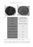
JOURNAL OF COSMETIC SCIENCE 118 hand-picked, and the seeds are removed from the pulp and red fi ber. Oil is obtained from the seeds by cold pressing. The coproduct of the oil extraction is the seedcake. The seed- cake is micronized and was used to perform the extraction. PREPARATION OF PLANT EXTRACT Baobab seedcake was fi rst ground with a blender to obtain a powder. Then, PSR technol- ogy patented process was applied to obtain the small RNA-enriched baobab extract and other phytochemicals. The extract was then diluted by 30% with either water or a cos- metic solvent such as glycerin. The extract was then transferred into sterile bottles and sterilized at low temperature. To obtain a plant extract placebo for the biological evalua- tion, another baobab extract that did not contain small RNAs was prepared. These two baobab extracts were comparatively studied for their effects on skin. PHYTOCHEMICAL ANALYSIS OF THE EXTRACT Phytochemical screening was performed on the baobab seedcake and on the fi nal extract to determine the dry weight and the quantifi cation of total protein and sugar. Polyphenol and amino acid content analysis was performed only on the fi nal extract. Dry weight of the raw material and of the fi nal extract was determined by placing 2 g of baobab seedcake powder in a metal cup in duplicate for 2 h at 105°C in a ventilated oven (Memmert, Schwabach, Germany). Protein content was determined by Lowry protein assay, which was used to quantify the total protein content of the extract (13). Protein measurement with the Folin phenol re- agent was used to quantify the total protein content of the extract. The Lowry method is based on the reaction of Cu+, produced by the oxidation of peptide bonds, with Folin– Ciocalteu reagent. The absorbance of the sample is read on the spectrophotometer at 550 nm. The protein content is determined using a BSA standard curve. Sugar content was determined colorimetrically via an adaptation of the assay described by Dubois et al. (14). This analysis consisted of concentrated sulfuric acid, which was then reacted with phenol to form a colored complex. The absorbance of the complex was read on the spectrophotometer at 490 nm. The sugar content was determined using a glucose standard curve. Polyphenolic compound quantity was determined using the Folin–Ciocalteu assay (15). Polyphenol compounds in the sample reacted with the Folin–Ciocalteu reagent, and the oxidation of the reagent turns the polyphenol compounds blue. The absorbance of the sample was read on the spectrophotometer at 760 nm. The content was expressed as gal- lic acid equivalents using a gallic acid standard curve. Amino acids were quantifi ed using a protocol published by Moore and Stein (16). The free amino acid content of the baobab extract was assessed by the formation of a colored complex, following the rupture of the amine and carboxylic functions by the reagent ninhydrin. The absorbance of the complex was read on the spectrophotometer at 570 nm. The total amino acids content was determined using a standard curve of amino acids pool. For the analysis of quality and integrity of the small RNAs present in our baobab extract, the Agilent 2100 Bioanalyzer system was used, with a small RNA Chip Kit according to the manufacturer’s instructions (Agilent, Santa Clara, CA).

PLANT SMALL RNA TECHNOLOGY 119 CELL CULTURE Fibroblasts were extracted from different normal human skin samples obtained from do- nors and were grown in Dulbecco’s modifi ed eagle medium (DMEM) 1 g/L glucose (Lonza, Verviers, Belgium) supplemented with 10% of FBS (Lonza), 2 mM of L-Glutamine (Lonza), and 100 μg/ml of Primocin (InvivoGen, San Diego, CA). Cell cultures were maintained at 37°C in a 5% CO2-humidifi ed atmosphere. In the model of replicative senescence, fi broblasts were cultured over a 3-mo period. At each passage, when cells reached confl uence, a part of these cells were frozen and the other part was maintained in culture. After different passages (P-X), the cells were thawed and treated, or not (control condition), overnight or for a period of 24 or 48 h with 1% PSR baobab extract or with 1% placebo (baobab extract without small RNAs) (twice a day). HUMAN SKIN SAMPLES Ex vivo skin samples were obtained from donors after undergoing abdominal plastic sur- gery. On skin biopsies of 6 mm in diameter, a topical application of 20 μl of either 1X phosphate-buffered saline (PBS) (control) or 1% PSR baobab extract or 1% placebo di- luted in 1X PBS (baobab extract without small RNAs) was applied twice a day for 48 h. Then, tissues were fi xed in formalin and embedded in molten paraffi n wax. qPCR After treatment, the culture medium was removed and the cells were rinsed with cold PBS. The mirVana miRNA Isolation Kit (Ambion, Naugatuck, CT) was used to extract total RNAs. Then, these RNAs were reverse-transcribed with the High-Capacity cDNA Reverse Transcription Kit with RNase inhibitors (Applied Biosystems, Foster City, CA). The reverse transcription was performed on 0.25 μg of total RNA using a MiniCycler thermocycler (MJ Research, Waltham, MA). Finally, real-time PCR was performed on a StepOnePlus thermocycler (Applied Biosystems). Collagen I, miRNA-19b or Drosha TaqMan Gene Expression Assay (Applied Biosystems) and TaqMan Gene Expression Master Mix (Applied Biosystems) were involved. RNU44 TaqMan Gene Expression As- say was used as an endogenous control to miRNA expression studies and 18S TaqMan Gene Expression Assay to Collagen I and Drosha expression study. The comparative Ct method was used for relative quantifi cation of target expressions (17), and the StepOne* Software (Applied Biosystems) was used for data treatment. SENESCENCE-ASSOCIATED (SA) β-GALACTOSIDASE STAINING Fibroblasts were washed in PBS, fi xed for 3 min in 2% formaldehyde and 0.2% glutaral- dehyde solution, washed, and incubated overnight at 37°C with SA β-galactosidase stain solution at 1 mg/ml [MgCl2 at 2 mM, K3Fe(CN)6 at 5 mM, K4Fe(CN)6 at 5 mM, NaCl at 150 mM, citric acid at 40 mM, and X-gal (Qiagen, Hilden, Germany)]. The slides were rinsed and mounted in Aquatex (Merck, Kenilworth, NJ). Detection was managed and examined using a Nikon Eclipse E600 microscope with a ×20 objective. Photos were taken with a QImaging* EXi blue camera and processed by using the Q-Capture Pro 7 (QImaging, Survey, BC, Canada) acquisition software.
Purchased for the exclusive use of nofirst nolast (unknown)
From: SCC Media Library & Resource Center (library.scconline.org)