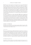
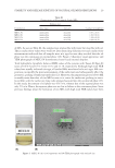
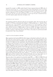
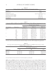
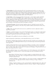
EXAMINING SKIN-BRIGHTENING INGREDIENTS USING APPLE SLICES 13 and is often used in in vitro studies to examine potential skin-brightening ingredients for topical applications (1). However, the enzyme tends to be expensive, and as noted earlier, the use of the enzyme in the kinetic in vitro study of tyrosinase inhibition is challenging because the study is kinetic (i.e., requires attention to time) and uses expensive spectro- photometric equipment that is not always available in skin care product formulating laboratories. In the recent work reported by Lin et al. (11,12), multiple scanning electrochemical microscopic mapping was used to examine tyrosinase activity in banana slices and in mushroom-derived tyrosinase directly, and in a later publication, the same technique was used to examine melanin in human skin melanoma biopsy samples. This work demon- strates the similarity between the enzymatic activity of tyrosinase in banana skins and in human skin. The work is intended to try and expand the use of electrochemical micro- scopic mapping as a means of detecting and staging melanoma tumors in human skin. We wish to report here efforts to use apple slices as a model for skin melanin production as a straightforward way to screen potential skin-brightening ingredients. We will discuss some parameters that were found to be important in using apple slices as a sub- strate to test skin-brightening ingredients. As will be shown, it does appear that this simple method could also potentially work to screen various types of skin-brightening ingredients including, potentially, fi nished formulations that are diffi cult to analyze with tissue equivalents or the in vitro tyrosinase assay. MATERIALS AND METHODS APPLE SLICES Apples used in the current study were purchased from a local supermarket and included two varieties, McIntosh apples and honey crisp apples. Initial work started by simply cutting the apples in half, but it was quickly realized that using a standard apple corer worked much better, Figure 1. The slicer allowed for the quick creation of up to eight slices, and, using only six appears adequate to obtain statistically signifi cant testing results using the chromameter. CHROMAMETER A DermaLab chromameter (Cortex Technologies) was used to measure the apple slice color. This instrument is well known for its use in skin melanin work (13). The proper placement of the chromameter probe on the apple slices is shown in Figure 2. The instru- ment measures in the CIE L*a*b* color space. The instrument also calculates an “erythema index” (a measure of redness) and a “melanin index” (a measure of brownness). As will be shown, for the purposes of examining darkening in apple slices, the erythema index appears to work best. Reasons for this will be discussed in more detail in the following paragraphs. The differences in the measurements for the single apple studies were analyzed for statistical signifi cance using the TTEST function in Excel (two-tailed and two-sample paired). The differences in the measurements for the multiple apple studies (azelaic acid study) were analyzed for statistical signifi cance using the TTEST function in Excel (two-tailed, two-sample unpaired unequal variance).

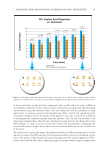
JOURNAL OF COSMETIC SCIENCE 14 AZELAIC ACID The source of azelaic acid used in the present study was Jeesperse® OptiDerm™ A-Z (Jeen International, Fairfi eld, NJ). The product is available as a water-dispersible powder con- taining 25% azelaic acid. When introduced into water at 10%, the Jeesperse® OptiDerm™ A-Z disperses to provide a white, low-viscosity emulsion containing 2.5% azelaic acid (Figure 3). RES ULTS AND DISCUSSION INI TIAL EXAMINATION OF VARIOUS KINDS OF APPLE SLICES Ini tial efforts to examine apple slices as potential melanin creation substrates began by examining simple slices of apples. It was found that in examining two initial apples using the chromameter, using the erythema index measurement, there was a rapid but predict- able increase in response from the surface of the apples as measured over a time course of 24 h. The McIntosh apple slices showed statistically signifi cant increases in the erythema index response over the course of 24 h (Figure 4). However, it was noted that the initial response was very rapid, happening within a 60-min time frame. This was a concern because it would mean that the treatment and measurement of the apple slices would have to take place very quickly to be able to detect a meaningful response. To take the measurements using the DermaLab chromameter, the device is placed on the surface of the apple and then on activation, the chromameter fl ashes two light-emitting diodes and records the resulting color response in a photodiode in the instrument head. Each apple slice is large enough to easily gather the four measurements per slice that the instrument Figure 1. Apple slicing apparatus.
Purchased for the exclusive use of nofirst nolast (unknown)
From: SCC Media Library & Resource Center (library.scconline.org)