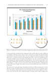
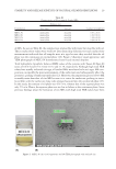
STABILITY AND RELEASE KINETICS OF NATURAL OIL MICROEMULSIONS 25 PREPARATI ON OF NICOTINAMIDE-LOADED MES Pseudoter nary phase diagrams of systems containing various ratios of Tween 80:Span 80, either virgin coconut oil or olive oil, and water were constructed by titration method. Tween 80 and Span 80 were studied at the S:CoS ratios of 1:0, 0.9:0.1, 0.8:0.2, 0.7:0.3, 0.6:0.4, 0.5:0.5, 0.4:0.6, 0.3:0.7, 0.2:0.8, and 0.1:0.9. Afterward, four formulations of blank MEs were selected from the middle points of four large ME regions in four pseudoternary phase diagrams. In this study, the chosen systems were composed of 0.7:0.3 or 0.6:0.4 Tween 80:Span 80 as the surfactant blend, virgin coconut oil or olive oil as the oil phase, and water as the aqueous phase. Blank MEs were prepared by simply mixing all components in the determined weight ratios. Afterward, 3% w/w nicotinamide-loaded MEs were prepared by mixing 3% w/w of nicotinamide powder and 97% w/w of each blank ME with a magnetic stirrer until nicotinamide powder completely dissolved. CHARACTER IZATION OF NICOTINAMIDE-LOADED MES The obtai ned nicotinamide-loaded MEs were left at room temperature (RT) overnight before being characterized. They were visually observed for appearance. They were investigated for isotropic property under polarized light microscope (Olympus BX61, Tokyo, Japan) to confi rm for ME formation. ME type was determined by drop dilution test into water as well as oil and by conductivity measurement with a conductivity meter (FiveEasy, Mettler Toledo, Greifensee, Switzerland). In case of drop dilution test into water, if the sample was miscible with water, it was defi ned as oil-in-water (o/w) because water was the external phase. By contrast, if the sample was immiscible with water, it was defi ned as w/o. In case of drop dilution test into oil, the results should be vice versa. The conductivity has been usually low in w/o, whereas high in o/w MEs (16). Transmission electron microscope (TEM, JEM-2010, JEOL, Tokyo, Japan) was used to illustrate the sample microstructure. Briefl y, a sample was dropped on a Formvar carbon fi lm on 200-mesh copper grid and left at RT until dry. Subsequently, the ob- tained sample was observed under TEM at magnifi cation of ×100,000. Viscosity val- ues were measured in triplicate by a rheometer (DV III Ultra Programmable Rheometer, Brookfi eld Engineering Laboratories, Middleboro, MA) using a spindle number SC4-31 with fi ve different shearing speeds at 32° ± 1°C. STABILITY EVA LUATION OF NICOTINAMIDE-LOADED MES Nicotinamide- loaded MEs were kept in clear glass containers at 4°C, RT, and 45°C for 3 mo. Their physical changes such as phase separation, turbidity, precipitation, and discolor- ation were observed every month. Their chemical stability was evaluated by analysis of amounts of nicotinamide remaining in the samples. Briefl y, an accurate weight (0.05 g) of each nicotinamide-loaded ME was vigorously mixed and diluted with isotonic phosphate buffer solution (PBS) pH 7.4 into an appropriate concentration. Afterward, each obtained sample was fi ltered by a syringe fi lter and analyzed for nicotinamide content by validated high-performance liquid chromatography (HPLC) technique. All experiments were per- formed in triplicate.

JOURNAL OF COSMETIC SCIENCE 26 IN VITRO RELEAS E STUDY OF NICOTINAMIDE-LOADED MES Two selected ni cotinamide-loaded MEs prepared with the same S:CoS but different oils were evaluated for in vitro release profi les and kinetics. The release study was performed by modifi ed Franz diffusion cells (Model 57-6 M, Hanson Research Corporation, Chatsworth, CA). A membrane model was dialysis membrane with the molar weight cutoff 3500 Dalton (Spectra/Por®3, Spectrum laboratories, New Brunswick, NJ). The membrane was cut into appropriate sizes and soaked in the receptor fl uid for 30 min before placed between donor and receptor chambers of the diffusion cell. Twelve milliliters of degassed PBS was added into each receptor chamber, stirred at a speed 200 rpm by a magnetic stirrer, and thermostatically maintained at 37° ± 0.5°C. An accurate weight (1 g) of the sample was applied on the membrane with the diffusion area of 1.77 cm2 in the donor chamber. At specifi ed time intervals (0.5, 1, 2, 4, 6, 8, 10, 12, and 24 h), 500 μL of receptor fl uid was withdrawn from the receptor chamber and immediately replaced with equal volume of PBS. The withdrawal samples were examined for nicotinamide concentrations by a validated HPLC technique. Experiments were carried out in triplicate for each formulation. The cumulative amount of released nicotinamide through the dialysis membrane into the receptor fl uid (Q, μg/cm2) was calculated by equation (1). Subsequently, the release data were further analyzed using three different kinetics models, i.e., zero order, fi rst order, and Higuchi model as exhibited in equations (2)–(4), respectively. 1 0 , t r t s i i Q V C V C (1) where Ct is the conc entr ation of nicotinamide in the receptor fl uid at each sampling time (t), Ci is the concentration of nicotinamide of the ith sample, and Vr and Vs are the volumes of the receptor fl uid and the sample, respectively. 0 0 Zeroorder : t Q Q k t (2) 0 Firstorder : ln lnQ t f Q k t (3) 1/2 Higuchi model :Q t H k t (4) where Qt is cumul ative amou nt of nicotinamide released in time (t), Q0 is initial amounts of nicotinamide in the formulations. The k0, kf, and kH are release rate constants of zero order, fi rst order, and Higuchi model, respectively. ANALYSIS OF NICOTINAMIDE T he HPLC technique was slightly modifi ed from previous studies (11,17). HPLC system (Agilent 1100 series, Palo Alto, CA) was used. Stationary phase was a reversed phase column (Luna® C18, 150 × 4.6 mm, 5 μm particle size, Phenomenex Inc., Torrance, CA). Mobile phase was a mixture of 0.1% triethylamine in 0.067 M monobasic potassium phosphate buffer pH 6.7 and acetonitrile (96.15:3.85 v/v). Mobile phase was fi ltered through the 0.45-μm nylon fi lter and degassed before use. Its fl ow rate was controlled at 1.0 mL/min. The injection
Purchased for the exclusive use of nofirst nolast (unknown)
From: SCC Media Library & Resource Center (library.scconline.org)